From: Structural Components and Architectures of RNA Exosomes
Copyright © 2000-2013, Landes Bioscience.

An official website of the United States government

NCBI Bookshelf. A service of the National Library of Medicine, National Institutes of Health.
Madame Curie Bioscience Database [Internet]. Austin (TX): Landes Bioscience; 2000-2013.

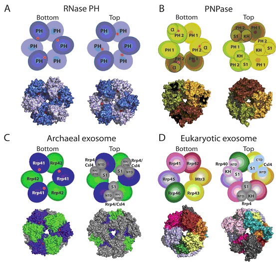
'Exosomes' from bacteria, archaea and eukaryotes have a similar architecture. RNase PH, PNPase, archaeal exosome and eukaryotic core exosome structures and schematics are depicted in two orientations which we denote bottom and top. Architectures emphasize a six-component ring with or without phosphorolytic active sites (shown as red dots in cartoon representation and red surfaces in the surface representations of the respective structures). A) RNase PH. The Aquifex aeolicus RNase PH structure (PDB ID = 1UDN) forms a homohexamer of PH subunits (colored dark blue and light blue, for clarity). B) PNPase. The S. antibioticus PNPase structure (PDB ID = 1E3P) forms a homotrimer. PNPase protomers are colored light yellow, dark yellow and light brown to distinguish the homotrimer of RNase PH 1-like (PH 1) and RNase PH 2-like (PH 2) domains. The α domain was omitted to enable visualization of the phosphorolytic active sites in the bottom view (red dots). C) Archaeal exosome. The S. solfataricus archaeal exosome (PDB ID = 2JE6) is depicted with Rrp41 subunits (blue) and Rrp42 subunits (green) which form the six-component ring. Top view in the schematic shows the orientation of Csl4 (N-Terminal Domain, S1 domain and C-Terminal Domain) and Rrp4 (N-Terminal Domain, S1 domain and KH domain) labeled and shown in grey. The surface representation of the structure depicts the Rrp4-bound archaeal exosome. Residues in the active site are partially occluded from view such that the active site appears as two discontinuous red surfaces. D) Eukaryotic core exosome. The eukaryotic exosome is shown from H. sapiens (PDB ID = 2NN6). Subunits Rrp41 (magenta), Rrp42 (red), Mtr3 (orange), Rrp43 (yellow), Rrp46 (green), Rrp45 (blue) form the six-component ring. Subunits Rrp40 (light pink), Csl4 (cyan) and Rrp4 (grey) form the three-component cap. No phosphorolytic active site exists in the eukaryotic core exosome.
From: Structural Components and Architectures of RNA Exosomes
Your browsing activity is empty.
Activity recording is turned off.
See more...