Continuing Education Activity
Low vision, visual impairment, and blindness are broad terms encompassing a range of conditions affecting an individual's ability to see and function in daily life. Low vision describes those whose vision cannot be fully corrected by glasses, contact lenses, refractive surgery, or other surgery. Visual impairment describes those whose decreased visual function interferes with the ability of one to perform their activities of daily living. However, the majority of causes of vision impairment can be prevented or rectified. As aging occurs, more and more blindness cases are added to the global burden, leading to an increased demand for eye healthcare services. The leading causes of blindness globally are cataracts, glaucoma, uncorrected refractive errors, age-related macular degeneration, and diabetic retinopathy. This activity provides an in-depth exploration of the various facets of blindness, low vision, and visual impairment, with a special emphasis on causes, legal interpretations, and the role of the interprofessional healthcare team in managing these patients.
Objectives:
Identify the etiology of blindness.
Assess vision loss based on patient evaluation.
Implement various treatment and management options available for blindness.
Explain interprofessional team strategies for improving coordination and outcomes in blind individuals.
Access free multiple choice questions on this topic.
Introduction
Total blindness, low vision, and visual impairment encompass a diverse range of conditions, each with its unique characteristics and implications for daily life.
Total blindness describes those who have a complete lack of light perception, documented as no light perception (NLP). Only about 15% of people with eye disorders have total blindness, and the majority of those with visual impairment have some level of vision.[1]
Low vision describes those whose vision cannot be fully corrected by conventional methods such as glasses, contact lenses, medicine, surgery, magnification aids, or assistive technology.[2]
Visual impairment describes those whose decreased visual function interferes with the ability of one to perform their activities of daily living, such as reading, driving, and watching TV. Visual impairment is defined based on function instead of using visual acuity or visual field cutoff values, and includes those who have low vision or who are blind.[3]
Symptoms of visual impairment include:
Legal blindness is defined by the United States Social Security Administration (SSA) to determine those who are eligible to receive disability benefits, tax exemption programs, and rehabilitation training. SSA uses visual acuity or visual field results to determine this eligibility.[5] A person is considered legally blind if he/she has central visual acuity of 20/200 or worse in the better-seeing eye with the best correction (using glasses or contact lenses) at a distance or if he/she has visual field restriction where the widest diameter is 20 degrees or less in the better-seeing eye.
There are several tests used to measure visual acuity or visual fields.[6][7] Visual acuity testing for distance is carried out using the Snellen visual acuity chart or another test that is comparable to the Snellen methodology. In 2007, SSA updated the criteria for measuring visual acuity by allowing newer low-vision test charts to be used instead of Snellen acuity charts alone. Under this update, if a person cannot read at least one letter on the 20/100 line, he or she will be classified as legally blind.[8]
Acceptable tests for visual field testing include automated static perimetry such as Humphrey Field Analyzer (HFA) 30-2, HFA 24-2, and Octopus 32, kinetic perimetry such as Goldmann perimetry or HFA "SSA Test Kinetic." Screening tests, including confrontation tests, tangent screen tests, and static screening tests, are not accepted forms of testing to determine legal blindness.
The World Health Organization (WHO) classifies visual impairment into the following categories based on visual acuity or a visual field of the better-seeing eye.
Normal: 20/10-20/25
Near normal visual impairment: 20/30-20/60
Moderate visual impairment: 20/70-20/160
Severe visual impairment: 20/200-20/400 or 11-20 degrees on visual field
Profound visual impairment: 20/500-20/1000 visual acuity or 6- 10 degrees on visual field
Near total visual impairment: Counting fingers, hand motion, light perception, or 5 degrees or less on visual field
Total visual impairment: No light perception
WHO also defines blindness as visual acuity of 3/60 (Snellen’s chart) or less or its equivalent (see Table. WHO and the National Programme on Control of Blindness [NPCB] classification of blindness). In the absence of visual acuity charts, when paramedical staff takes the visual acuity, it is defined as the inability to count fingers at a distance of 3 meters in daylight to indicate 3/60 or its equivalent.[9]
Table. WHO and the National Programme on Control of Blindness (NPCB) classification of blindness [1].
Etiology
There can be many different causes of blindness. The leading causes of blindness worldwide are cataracts, age-related macular degeneration, glaucoma, diabetic retinopathy, and trachoma.[10]
Cataracts: A condition that causes yellowing and hardening of the lens in the eye; the leading cause of blindness worldwide in developing and developed countries
Age-related macular degeneration: A condition that damages a part of the retina known as the macula; the leading cause of blindness in Caucasians and those aged 65 and older
Glaucoma: A condition that damages the optic nerve; the leading cause of blindness in African Americans
Diabetic retinopathy: A condition due to systemic diabetes; the leading cause of new blindness in adults between 25 and 64
Trachoma: A condition caused by the
Chlamydia trachomatis bacteria
; incidence is decreasing due to public health action
[11]
The number of persons with reduced vision due to uncorrected refractive error exceeds that of those with the conditions mentioned above. However, uncorrected refractive errors can be easily treated or "cured" with proper vision correction.
Estimates are that approximately 90% of the visually impaired population lives in developing countries or low-income circumstances. About 80% of all visual impairments worldwide can be prevented, treated, or cured with proper eye care.
The most common causes of blindness are:
Cataracts (51%)
Glaucoma (8%)
Age-related macular degeneration (5%)
Corneal opacification (4%)
Childhood blindness (4%)
Refractive errors (3%)
Trachoma (3%)
Diabetic retinopathy (1%)
Undetermined (21%)
In developed countries, the major causes of blindness are:
In children, the major causes of blindness are:
Xerophthalmia
Congenital glaucoma
Congenital cataract
Optic atrophy
Trauma
Amblyopia
Refractive errors
Epidemiology
According to WHO, approximately 285 million people have a visual impairment, including 39 million who are blind and 246 million who have low vision. Of those who are blind, 90% live in developing countries. For each blind person worldwide, an average of 3.4 people have low vision, with the country and regional variation ranging from 2.4 to 5.5 people.[10][13] According to another report, 180 million people worldwide are visually disabled, including 45 million who are blind (80% of this blindness is preventable).[14]
More than 82% of all people who are blind are 50 years of age and older. Due to the expected number of years lived in blindness, childhood blindness is a significant problem, with an estimated 1.4 million children under age 15 who are blind. Females have a significantly higher risk of being visually impaired than males.[15]
Approximate prevalence of blindness due to various causes:
A sizeable portion of the blind population (32%) is between 40 and 70 years old, and 60% is over 60 years old. The yearly incidence of blindness due to cataracts is 2 million cases. According to the national survey analysis on blindness 2001-2002, the estimated prevalence of blindness is 8.5% in the over-50 age group and 1.1% in the general population. Approximately 7% of children are suffering from ocular conditions leading to low vision and problems with eyesight.[17]
Some of the ocular conditions leading to blindness are more common in children, such as refractive error, trachoma, conjunctivitis, and malnourishment.[18] The ocular conditions more common in women are cataracts, conjunctivitis, and trachoma, leading to a greater prevalence of blindness in women. Occupation-related blindness is more common in factory workers, workshops, and industrial workers who are more prone to penetrating and perforating injuries. These people are more exposed to dust, flying objects and particles, radiation, fumes, and gases.[19] As per previous studies, it has been documented that blindness is more common in poor socioeconomic strata, people with low education, poor patients, and patients with low standards of personal and community hygiene.[20]
Pathophysiology
Conditions causing visual impairment vary widely and can be genetic, congenital, or acquired. Vision loss can occur gradually or suddenly and can result in central vision loss, peripheral vision loss, overall blur, a decrease in contrast sensitivity, color vision difficulties, night vision blindness, and glare or light sensitivity issues. The cause of vision loss determines whether it involves one type alone or a combination of presentations.[21][22]
History and Physical
Performing a thorough case history is essential. The case history should include:
Patient's visual and ocular history
Family visual and ocular history
Patient medical history, including medications
Family medical history
Social history
Vocational, educational, and hobby history
[23]
Subsequently, an ocular examination is necessary to assess the patient's current visual status and function; it should include:
Visual acuity (distance and near)
Refraction
Pupils, motility, and binocular vision
Visual fields (central and peripheral fields of vision)
Glare testing, color vision, contrast sensitivity, binocular single vision
Gross examination on torch light, including ocular movements
Detailed anterior segment examination
Detailed posterior segment examination
Tonometry (intraocular pressure measurement)
Evaluation
An ocular health examination should typically include visual acuity, visual field, extraocular muscles, pupil, binocular vision testing, intraocular pressure, anterior segment, and posterior segment evaluation with dilated fundus exams. Supplemental testing such as formalized visual field testing, color vision tests, contrast sensitivity tests, visual evoked potential, electroretinography, electrooculography, optical coherence tomography, fluorescein angiography, and genetic testing may be necessary to help determine the etiology and to monitor and properly manage the condition.[25]
Treatment / Management
Comprehensive eye examinations are necessary to prevent, detect, treat, and manage the ocular conditions that can lead to blindness. Eye exams should include ocular health exams and not just vision and sight checks for glasses or contact lens prescriptions.[26]
Many international, national, and local organizations are working to bring more awareness about vision and eye health, provide access to healthcare, and fight blindness. Research is being done worldwide to study the etiologies of many eye conditions causing permanent vision loss and to develop a proper treatment to control or cure these conditions. WHO is the leader in monitoring trends, raising awareness, and coordinating efforts to fight blindness.[27]
Low-vision rehabilitation services are available to help patients maximize their remaining vision, maintain their independence, and improve their quality of life.[28] These services are provided by a multidisciplinary team of low-vision professionals, including low-vision doctors (optometrists or ophthalmologists), low-vision therapists, occupational therapists, rehabilitation teachers, orientation and mobility specialists, vocational rehabilitation specialists, social workers, and other rehabilitation low-vision professionals. Please refer to the review article titled "Low Vision Rehabilitation" for additional information.[29]
As determined by NPCB, there are various levels at which blindness is controlled.
Primary Assessment
Primary Assessment is performed by prevalence surveys. It is necessary to assess the magnitude, geographical distribution of pathology, and etiology of blindness within a particular town, place, or country.[30]
Intervention
Primary Eye Care
Primary eye care is the management of eye conditions at the grassroots level by trained workers. The paramedical staff is the first to make contact with people in the community. The staff is trained and dedicated to referring needy patients to the nearest primary health center and district hospitals. They also help promote practices such as basic hygiene, sanitation, dietary management, and the safety of patients. The main aim is to improve the quality of eyecare by primary eye care approach and improve the quality of present resources.[31]
Secondary Eye Care
This involves the management of common ocular conditions such as cataracts, trichiasis, glaucoma, ocular trauma, and entropion. Secondary eye care is taken care of at primary healthcare centers and district hospitals where there are well-developed eye departments. A strong component of secondary eye care is mobile eye clinics. This strategy's main advantage is treating problem-specific concerns, utilizing local resources, and providing cost-effective eye care to the population at a peripheral level.[32]
Tertiary Eye Care
This level of eyecare is provided at the national and regional capitals and is provided at the level of medical colleges and medical institutes. The ocular pathologies that can be managed at this level are retinal surgeries in retinal detachment, corneal transplant, trabeculectomy, orbitotomy, etc, which are not available at secondary centers. The other services offered are the education of blind people and service utilization for creating employment. The various special programs offered for disease-specific control are trachoma control, xerophthalmia measures with vitamin A prophylaxis, school screening, health education, and occupational eye services.[33]
Long-term Outcomes
To improve quality of life, hygiene, nutrition, personal hygiene, and sanitation measures must be taken into consideration.[34]
Differential Diagnosis
Glaucoma Causes
Acute angle-closure glaucoma
Primary open-angle glaucoma
[35]Primary angle-closure glaucoma
Normal tension glaucoma
Neovascular glaucoma
Malignant glaucoma
Pseudoexfoliation glaucoma
Inflammatory glaucoma
Lens-induced glaucoma
Traumatic glaucoma
Iridocorneal endothelial syndrome
Glaucoma associated with intraocular tumors
Glaucoma secondary to epithelial ingrowth
Primary congenital glaucoma
Iridocorneal dysgenesis
Glaucoma in phacomatoses
Corneal Causes
Fungal keratitis
Bacterial keratitis
Viral keratitis
Herpes zoster ophthalmicus
Interstitial keratitis
Protozoan keratitis
Corneal dystrophies
Graft rejection
Helminthic keratitis
Bacterial hypersensitivity-mediated corneal diseases
Rosacea keratitis
Peripheral ulcerative keratitis
Mooren’s ulcer
Penetrating and perforating corneal injuries
Keratoconus
Keratoglobus
Pellucid marginal degeneration
Exposure keratopathy
Congenital anomalies of the cornea
Neurotrophic keratitis
Chemical injury
Thermal injury
Retinal Causes
Endophthalmitis
Diabetic retinopathy
Nondiabetic retinopathy
Central and branch retinal venous occlusion
Central retinal arterial occlusion
Ocular ischaemic syndrome
Hypertensive retinopathy
Sickle-cell retinopathy
Thalassaemia retinopathy
Retinopathy of prematurity
Retinal artery macroaneurysm
Primary retinal telangiectasia
Eales disease
Radiation retinopathy
Purtscher retinopathy
Valsalva retinopathy
Lipaemia retinalis
Retinopathy in blood disorders
Retinal detachment
Intraretinal foreign body
Vitreous hemorrhage
Uveal Causes
Anterior uveitis
Intermediate uveitis
Posterior uveitis
Panuveitis
Uveitis in spondyloarthropathies
Fuchs uveitis syndrome
Uveitis in juvenile idiopathic arthritis
Uveitis in bowel disease
Uveitis in renal disease
Intermediate uveitis
Vogt–Koyanagi–Harada (VKH) syndrome
Sympathetic ophthalmitis
Lens-induced uveitis
Sarcoidosis
Behçet disease
Parasitic uveitis
Viral uveitis
Fungal uveitis
Bacterial uveitis
Sclerokeratouveitis
Macular Causes
Age-related macular degeneration
Angioid streaks
Choroidal folds
Hypotony maculopathy
Solar retinopathy
Focal choroidal excavation
Retinal angiomatous proliferation
Polypoidal choroidal vasculopathy
Peripheral exudative haemorrhagic
Chorioretinopathy
Idiopathic choroidal neovascularization
Vitreomacular interface disorders
Central serous chorioretinopathy
Idiopathic macular telangiectasia
Cystoid macular edema
Microcystic macular edema
Degenerative myopia
Neuro-ophthalmic Causes
Giant-cell arteritis
Optic neuritis
Papilledema
Retrobulbar neuritis
Orbital apex syndrome
Arteritic ischemic optic neuropathy
Nonarteritic ischemic optic neuropathy
Cranial nerve palsy
Tolosa-Hunt syndrome
Foster Kennedy syndrome
Pseudo–Tolosa-Hunt syndrome
Toxic optic neuropathy
Nutritional optic neuropathy
Orbital Causes
Prognosis
Depending on the condition, vision can be stable or progressive in nature. Conditions such as age-related macular degeneration only cause central vision loss and will not result in total blindness, whereas retinitis pigmentosa can cause peripheral vision loss leading to central vision loss and total blindness. Conditions causing blindness may be prevented with early detection, proper diagnosis, and treatment. It is recommended that people of all ages have a comprehensive eye examination every 1 to 2 years. For infants, InfantSEE is a free public health program available to provide comprehensive infant vision and eye health assessment within the first year of the infant's life.[40]
Complications
Complications that can cause blindness include:
Corneal perforation
Corneal abscess
Corneal melt
Lens dislocation
Lens abscess
Retinal detachment
Giant retinal tear
Endophthalmitis
Macular scar
Retinal sclopetaria
Chorioretinitis
Choroidal rupture
Choroidal detachment
Optic disc avulsion
Consultations
Any patient with sudden onset loss of vision should be evaluated by an ophthalmologist to rule out the cause of blindness. A meticulous ophthalmic examination is warranted in each to pinpoint the ocular pathology. Appropriate specialist consultation should be sought after screening if needed.[41]
Deterrence and Patient Education
A patient who is legally blind should be educated on the need for regular follow-up to prevent the development of further complications, as well as what measures can be taken to improve their lifestyle. A person with ocular disability and blindness should be advised that he or she is entitled to receive the ocular disability certificate and can apply for government benefits.[42]
Pearls and Other Issues
Blindness has affected millions worldwide, and it is the duty of ophthalmologists and paramedical staff to reduce this burden. A person with an ocular disability, if managed on time, can be prevented from developing irregular ocular sequelae. The cause of blindness should be ruled out in each case.[16]
Enhancing Healthcare Team Outcomes
Blindness in all its forms and severities confers serious morbidity to those affected, whether total blindness or visual impairments that include transient blindness or partial loss of field of vision. The assessment, diagnosis, and management of vision loss requires an interprofessional team approach, including physicians, specialists, specialty-trained nurses, optometrists, and pharmacists, all collaborating across disciplines to achieve optimal patient results.[43]
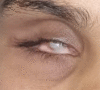
Digital image of the patient depicting leucomatous corneal opacity occupying the visual axis causing reduction in visual acuity Contributed by Bharat Gurnani, MD
Digital image of the patient depicting traumatic ptosis along with total corneal opacity post infective keratitis in a young patient Contributed by Bharat Gurnani, MD
References
- 1.
Vashist P, Senjam SS, Gupta V, Gupta N, Kumar A. Definition of blindness under National Programme for Control of Blindness: Do we need to revise it?
Indian J Ophthalmol. 2017 Feb;65(2):92-96. [
PMC free article: PMC5381306] [
PubMed: 28345562]
- 2.
Shah P, Schwartz SG, Gartner S, Scott IU, Flynn HW. Low vision services: a practical guide for the clinician.
Ther Adv Ophthalmol. 2018 Jan-Dec;10:2515841418776264. [
PMC free article: PMC6024512] [
PubMed: 29998224]
- 3.
Şahlı E, İdil A. A Common Approach to Low Vision: Examination and Rehabilitation of the Patient with Low Vision.
Turk J Ophthalmol. 2019 Apr 30;49(2):89-98. [
PMC free article: PMC6517854] [
PubMed: 31055894]
- 4.
Hu CX, Zangalli C, Hsieh M, Gupta L, Williams AL, Richman J, Spaeth GL. What do patients with glaucoma see? Visual symptoms reported by patients with glaucoma.
Am J Med Sci. 2014 Nov;348(5):403-9. [
PMC free article: PMC4206382] [
PubMed: 24992392]
- 5.
National Research Council (US) Committee on Disability Determination for Individuals with Visual Impairments.
Visual Impairments: Determining Eligibility for Social Security Benefits. Lennie P, Van Hemel SB, editors. National Academies Press (US); Washington (DC): 2002. [
PubMed: 25032291]
- 6.
Chakravarthy U, Bailey CC, Johnston RL, McKibbin M, Khan RS, Mahmood S, Downey L, Dhingra N, Brand C, Brittain CJ, Willis JR, Rabhi S, Muthutantri A, Cantrell RA. Characterizing Disease Burden and Progression of Geographic Atrophy Secondary to Age-Related Macular Degeneration.
Ophthalmology. 2018 Jun;125(6):842-849. [
PubMed: 29366564]
- 7.
Larsen PP, Thiele S, Krohne TU, Ziemssen F, Krummenauer F, Holz FG, Finger RP., OVIS-Study Group. Visual impairment and blindness in institutionalized elderly in Germany.
Graefes Arch Clin Exp Ophthalmol. 2019 Feb;257(2):363-370. [
PubMed: 30483949]
- 8.
Caltrider D, Gupta A, Tripathy K.
StatPearls [Internet]. StatPearls Publishing; Treasure Island (FL): May 1, 2024. Evaluation of Visual Acuity. [
PubMed: 33231977]
- 9.
Dandona L, Dandona R. Revision of visual impairment definitions in the International Statistical Classification of Diseases.
BMC Med. 2006 Mar 16;4:7. [
PMC free article: PMC1435919] [
PubMed: 16539739]
- 10.
Wolfram C, Schuster AK, Elflein HM, Nickels S, Schulz A, Wild PS, Beutel ME, Blettner M, Münzel T, Lackner KJ, Pfeiffer N. The Prevalence of Visual Impairment in the Adult Population.
Dtsch Arztebl Int. 2019 Apr 26;116(17):289-295. [
PMC free article: PMC6584831] [
PubMed: 31196384]
- 11.
Foster A, Resnikoff S. The impact of Vision 2020 on global blindness.
Eye (Lond). 2005 Oct;19(10):1133-5. [
PubMed: 16304595]
- 12.
Pascolini D, Mariotti SP. Global estimates of visual impairment: 2010.
Br J Ophthalmol. 2012 May;96(5):614-8. [
PubMed: 22133988]
- 13.
Hussain AHME, Ferdoush J, Mashreky SR, Rahman AKMF, Ferdausi N, Dalal K. Epidemiology of childhood blindness: A community-based study in Bangladesh.
PLoS One. 2019;14(6):e0211991. [
PMC free article: PMC6555501] [
PubMed: 31173584]
- 14.
Ackland P, Resnikoff S, Bourne R. World blindness and visual impairment: despite many successes, the problem is growing.
Community Eye Health. 2017;30(100):71-73. [
PMC free article: PMC5820628] [
PubMed: 29483748]
- 15.
Wadhwani M, Vashist P, Singh SS, Gupta V, Gupta N, Saxena R. Prevalence and causes of childhood blindness in India: A systematic review.
Indian J Ophthalmol. 2020 Feb;68(2):311-315. [
PMC free article: PMC7003592] [
PubMed: 31957718]
- 16.
Vashist P, Senjam SS, Gupta V, Gupta N, Shamanna BR, Wadhwani M, Shukla P, Manna S, Yadav S, Bharadwaj A. Blindness and visual impairment and their causes in India: Results of a nationally representative survey.
PLoS One. 2022;17(7):e0271736. [
PMC free article: PMC9302795] [
PubMed: 35862402]
- 17.
GBD 2019 Blindness and Vision Impairment Collaborators; Vision Loss Expert Group of the Global Burden of Disease Study. Causes of blindness and vision impairment in 2020 and trends over 30 years, and prevalence of avoidable blindness in relation to VISION 2020: the Right to Sight: an analysis for the Global Burden of Disease Study.
Lancet Glob Health. 2021 Feb;9(2):e144-e160. [
PMC free article: PMC7820391] [
PubMed: 33275949]
- 18.
Rao GN, Sabnam S, Pal S, Rizwan H, Thakur B, Pal A. Prevalence of ocular morbidity among children aged 17 years or younger in the eastern India.
Clin Ophthalmol. 2018;12:1645-1652. [
PMC free article: PMC6134961] [
PubMed: 30233126]
- 19.
Courtright P, West SK. Contribution of sex-linked biology and gender roles to disparities with trachoma.
Emerg Infect Dis. 2004 Nov;10(11):2012-6. [
PMC free article: PMC3328994] [
PubMed: 15550216]
- 20.
Habtamu E, Wondie T, Aweke S, Tadesse Z, Zerihun M, Zewdie Z, Callahan K, Emerson PM, Kuper H, Bailey RL, Mabey DC, Rajak SN, Polack S, Weiss HA, Burton MJ. Trachoma and Relative Poverty: A Case-Control Study.
PLoS Negl Trop Dis. 2015 Nov;9(11):e0004228. [
PMC free article: PMC4657919] [
PubMed: 26600211]
- 21.
Lozada KN, Cleveland PW, Smith JE. Orbital Trauma.
Semin Plast Surg. 2019 May;33(2):106-113. [
PMC free article: PMC6486387] [
PubMed: 31037047]
- 22.
Avogaro A, Fadini GP. Microvascular complications in diabetes: A growing concern for cardiologists.
Int J Cardiol. 2019 Sep 15;291:29-35. [
PubMed: 30833106]
- 23.
Takusewanya M. How to take a complete eye history.
Community Eye Health. 2019;32(107):44-45. [
PMC free article: PMC7041835] [
PubMed: 32123468]
- 24.
Musch DC, Niziol LM, Gillespie BW, Lichter PR, Janz NK. Binocular Measures of Visual Acuity and Visual Field versus Binocular Approximations.
Ophthalmology. 2017 Jul;124(7):1031-1038. [
PMC free article: PMC5483206] [
PubMed: 28408039]
- 25.
Moshirfar M, Murri MS, Shah TJ, Skanchy DF, Tuckfield JQ, Ronquillo YC, Birdsong OC, Hofstedt D, Hoopes PC. A Review of Corneal Endotheliitis and Endotheliopathy: Differential Diagnosis, Evaluation, and Treatment.
Ophthalmol Ther. 2019 Jun;8(2):195-213. [
PMC free article: PMC6514041] [
PubMed: 30859513]
- 26.
Yadav S, Tandon R. Comprehensive eye examination: what does it mean?
Community Eye Health. 2019;32(107):S1-S4. [
PMC free article: PMC7041818] [
PubMed: 32123482]
- 27.
Shrestha GS, Sigdel R, Shrestha JB, Sharma AK, Shrestha R, Mishra SK, Joshi SN. Awareness of Eye Health and Diseases among the Population of the Hilly Region of Nepal.
J Ophthalmic Vis Res. 2018 Oct-Dec;13(4):461-469. [
PMC free article: PMC6210871] [
PubMed: 30479718]
- 28.
Wang BZ, Pesudovs K, Keane MC, Daly A, Chen CS. Evaluating the effectiveness of multidisciplinary low-vision rehabilitation.
Optom Vis Sci. 2012 Sep;89(9):1399-408. [
PubMed: 22902419]
- 29.
Owsley C, McGwin G, Lee PP, Wasserman N, Searcey K. Characteristics of low-vision rehabilitation services in the United States.
Arch Ophthalmol. 2009 May;127(5):681-9. [
PMC free article: PMC2737181] [
PubMed: 19433720]
- 30.
Verma R, Khanna P, Prinja S, Rajput M, Arora V. The national programme for control of blindness in India.
Australas Med J. 2011;4(1):1-3. [
PMC free article: PMC3562965] [
PubMed: 23393496]
- 31.
Khan MA, Soni M, Khan MD. Development of primary eye care as an integrated part of comprehensive health care.
Community Eye Health. 1998;11(26):24-6. [
PMC free article: PMC1706055] [
PubMed: 17492028]
- 32.
Patel D. The role of secondary level eye care teams in engaging with and supporting primary eye care.
Community Eye Health. 2021;34(113):77-78. [
PMC free article: PMC9412127] [
PubMed: 36033412]
- 33.
Al Motowa S, Khandekar R, Al-Towerki A. Resources for eye care at secondary and tertiary level government institutions in Saudi Arabia.
Middle East Afr J Ophthalmol. 2014 Apr-Jun;21(2):142-6. [
PMC free article: PMC4005178] [
PubMed: 24791105]
- 34.
Muhoozi GKM, Atukunda P, Diep LM, Mwadime R, Kaaya AN, Skaare AB, Willumsen T, Westerberg AC, Iversen PO. Nutrition, hygiene, and stimulation education to improve growth, cognitive, language, and motor development among infants in Uganda: A cluster-randomized trial.
Matern Child Nutr. 2018 Apr;14(2):e12527. [
PMC free article: PMC6866193] [
PubMed: 28925580]
- 35.
Gurnani B, Srinivasan K, Venkatesh R, Kaur K. Do motivational cards really benefit sibling screening of primary open-angle glaucoma probands?
Indian J Ophthalmol. 2022 Dec;70(12):4158-4163. [
PMC free article: PMC9940586] [
PubMed: 36453305]
- 36.
Gurnani B, Christy J, Narayana S, Rajkumar P, Kaur K, Gubert J. Retrospective multifactorial analysis of
Pythium keratitis and review of literature.
Indian J Ophthalmol. 2021 May;69(5):1095-1101. [
PMC free article: PMC8186601] [
PubMed: 33913840]
- 37.
Gurnani B, Narayana S, Christy J, Rajkumar P, Kaur K, Gubert J. Successful management of pediatric pythium insidiosum keratitis with cyanoacrylate glue, linezolid, and azithromycin: Rare case report.
Eur J Ophthalmol. 2022 Sep;32(5):NP87-NP91. [
PubMed: 33779337]
- 38.
Gurnani B, Kaur K, Venugopal A, Srinivasan B, Bagga B, Iyer G, Christy J, Prajna L, Vanathi M, Garg P, Narayana S, Agarwal S, Sahu S.
Pythium insidiosum keratitis - A review.
Indian J Ophthalmol. 2022 Apr;70(4):1107-1120. [
PMC free article: PMC9240499] [
PubMed: 35325996]
- 39.
Gurnani B, Kaur K, Agarwal S, Lalgudi VG, Shekhawat NS, Venugopal A, Tripathy K, Srinivasan B, Iyer G, Gubert J. Pythium insidiosum Keratitis: Past, Present, and Future.
Ophthalmol Ther. 2022 Oct;11(5):1629-1653. [
PMC free article: PMC9255487] [
PubMed: 35788551]
- 40.
Sabel BA, Wang J, Cárdenas-Morales L, Faiq M, Heim C. Mental stress as consequence and cause of vision loss: the dawn of psychosomatic ophthalmology for preventive and personalized medicine.
EPMA J. 2018 Jun;9(2):133-160. [
PMC free article: PMC5972137] [
PubMed: 29896314]
- 41.
Prasad S, Galetta SL. Approach to the patient with acute monocular visual loss.
Neurol Clin Pract. 2012 Mar;2(1):14-23. [
PMC free article: PMC5766020] [
PubMed: 29443297]
- 42.
Stevens S. Assisting the blind and visually impaired: guidelines for eye health workers and other helpers.
Community Eye Health. 2003;16(45):7-9. [
PMC free article: PMC1705868] [
PubMed: 17491861]
- 43.
Demmin DL, Silverstein SM. Visual Impairment and Mental Health: Unmet Needs and Treatment Options.
Clin Ophthalmol. 2020;14:4229-4251. [
PMC free article: PMC7721280] [
PubMed: 33299297]
Disclosure: So Yeon Lee declares no relevant financial relationships with ineligible companies.
Disclosure: Bharat Gurnani declares no relevant financial relationships with ineligible companies.
Disclosure: Fassil Mesfin declares no relevant financial relationships with ineligible companies.