Focal and Generalized Slowing and Significance
EEG can provide evidence for underlying diffuse or focal cerebral dysfunction through demonstration of background slowing. The two main types of slowing are focal and generalized slowing. As previously discussed, generalized background slowing in the theta and delta frequency ranges is a normal finding on EEG when it represents developmental slowing in children, adolescents, and some young adults or the evolution of drowsiness and sleep activity. However, when there is intermittent or persistent focal slowing seen consistently over one head region, or persistent, unvarying, and unreactive focal or generalized slow wave activity in a vigilant adult patient, this slow wave activity should be considered pathologic and indicates corresponding focal or generalized cerebral dysfunction or both.
Focal Slowing
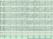
Focal slow wave activity on the EEG is indicative of focal cerebral pathology of the underlying brain region. Slowing may be intermittent or persistent, with more persistent or consistently slower activity generally indicating more severe underlying focal cerebral dysfunction. A variety of etiologies for focal cerebral dysfunction may be seen. When intermittent, focal slowing may indicate unveiling of subtle focal cerebral dysfunction owing to the effects of a sedating or hypnotic medication, although usually medication-induced slowing is generalized in nature. Focal brain lesions of a variety of causes to cortex, underlying white matter, or both may induce focal slowing. The various causes are too numerous to be comprehensive, but common examples include transient or permanent ischemia resulting from stroke, brain hemorrhage, tumors, traumatic injury, malformations of cortical development, nonstructural focal cerebral dysfunction corresponding to a focal epileptic focus, focal involvement of the cortex by neurodegeneration, arteriovenous malformations, and focal brain infection caused by bacterial cerebritis or viral encephalitis. See for an example of focal temporal regional slowing, which also shows a “breach rhythm,” with focally elevated background amplitude as a result of a skull defect and previous surgery in this area.
Focal slowing over the right temporal region as the result of a right temporal brain tumor in a 35-year-old man. Note the focal delta frequency slowing in the right temporal region as compared with the homologous normal right temporal region. Longitudinal (more...)
Generalized Slowing
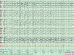
Generalized background slowing indicates diffuse cerebral dysfunction, which, similar to focal slowing, is also not specific as to cause. Several different etiologies may provoke generalized background slowing, including the effects of sedative centrally acting medications, neurodegenerative disorders, a widespread neurodevelopmental process, hydrocephalus, metabolic or toxic encephalopathy, CNS infectious disorders such as meningoencephalitis, or even a focal midline structural lesion involving deep midline brainstem, diencephalic structures, or both (producing a phenomenon known in older EEG literature as a “distance or projected rhythm,” which often selectively involves the anterior or bifrontal areas and produces a pattern known as frontal intermittent rhythmic delta activity, or FIRDA). See for an example of generalized EEG slowing resulting from a metabolic encephalopathy. Specific examples of clinical phenomena associated with generalized slowing on the EEG are discussed below.
FIRDA pattern with a slightly slower theta EEG background in an elderly man with a metabolic encephalopathy. Periods of well-formed posteriorly dominant alpha activity are still also seen. Longitudinal bipolar montage. Copyright 2013. Mayo Foundation (more...)
Encephalopathy/Delirium
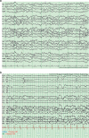
Delirium, also known as encephalopathy, is a reversible generalized confusional state induced by a systemic disorder. The clinical phenomena of confusion in a delirious state may closely resemble a complex partial or atypical absence seizure, involving blank staring with disorientation, inattention, and variable responsiveness, stupor with reduced vigilance, and unusual movements including myoclonic jerks. Encephalopathic patients may have acute symptomatic seizures, resulting in diagnostic confusion. EEG in a delirious patient may show either diffuse nonspecific nonepileptiform background slowing, or even epileptiform-appearing patterns such as triphasic waveforms (see for triphasic wave pattern), which are most common in patients with underlying associated hepatic or renal impairment or both, and resultant encephalopathy, although similar patterns may be induced by drug intoxication or adverse effects or other nonlesional causes of severe generalized cerebral dysfunction.
Generalized triphasic wave pattern and slowing in a 72-year-old man with hepatic encephalopathy. Longitudinal bipolar montage. Waveforms show characteristic anterior to posterior lag (see waveform marked by black arrow). Copyright 2013. Mayo Foundation (more...)
A distinctive pattern, which may be seen in some encephalopathic or even comatose patients, is lateralized periodic discharges (LPDs, formerly known as PLEDS), which may be focal, unilateral hemispheric, or even bilateral and independent. While LPDs are consistent with a heightened epileptogenic potential and may accompany patients having seizures (or even occur as a postictal and, somewhat controversially, on occasion as an ictal seizure pattern itself in prolonged status epilepticus), they may occur in a patient who is not having seizures when there is an acute destructive process involving the gray-white matter junction, especially in the context of herpes simplex virus (HSV) encephalitis, or following massive hemispheric ischemic infarction. See for an example of LPDs in HSV encephalitis.
LPDs over the left temporal region in a 75-year-old encephalopathic man with acute HSV encephalitis. Similar findings can be seen following acute ischemic infarction. Copyright 2013. Mayo Foundation for Medical Education and Research. All rights reserved. (more...)
Dementias
Early in the course of a slowly progressive neurodegenerative dementia, such as Alzheimer disease, the EEG may be normal during wakefulness and sleep. Later, as the disease progresses, there is frequently mild generalized background slowing. Focal slowing may also be seen.
A distinctive finding in a subacute, progressive dementia caused by prion disease (Creutzfeldt-Jakob disease, CJD) is the presence of periodic sharp-wave activity. While the EEG may be normal early in the course of CJD, by approximately the second to third month of symptoms, especially at the point of evolution of clinically overt myoclonus, periodic sharps appear, at times subtle or confined to a focal region (especially posteriorly); but with full-blown dementia and myoclonus, periodic sharp waves of 1- to 2-Hz frequency appear in a generalized distribution (see below).
Periodic sharp wave complexes in Creutzfeldt-Jakob disease. Generalized periodic sharp waves are seen in a 65-year-old man with rapidly evolving dementia and myoclonus later proven to have CJD. Longitudinal bipolar montage. This recording was obtained (more...)
Coma
Coma is a clinical state of eyes closed, irreversible unresponsiveness (at least temporarily), as opposed to sleep in which the unresponsive state is readily reversible to wakefulness. The hallmark of coma patterns is their lack of variability and relative (or absolute) lack of reactivity. Reactivity of the background (background frequency speeding up, or changing in reaction to physical or auditory stimuli) is a sign of relative integrity and considered relatively more favorable.
Several common EEG coma patterns have been described. The two patterns that are considered to have the worst prognosis for recovery following anoxic-ischemic encephalopathy are burst suppression (see ) and alpha coma, with other patterns considered intermediate (theta coma, ) or even favorable (spindle coma). However, prognostication should certainly not rely upon EEG alone, as the findings must be integrated into the clinical context and other ancillary tests such as neuroimaging.
Burst-suppression coma pattern following anoxic-ischemic brain injury as a result of cardiopulmonary arrest in a 58-year-old man with status myoclonus. Note the intervals of spike, polyspike, and slow wave discharges with intervening suppressed background (more...)
Theta coma pattern following heroin overdose in a 44-year-old comatose man post cardiac arrest. Note the relatively invariant theta and delta frequency slowing (predominantly theta) in a generalized distribution and lack of spontaneous variability in (more...)
Anesthetic Patterns
The EEG is often useful during surgical procedures to follow the depth of anesthesia and is particularly useful during neurovascular surgeries in which there is risk for thromboembolism and otherwise covert occurrence of cerebrovascular ischemia, such as carotid endarterectomy surgeries. below demonstrates a typical anesthetic pattern, characterized by predominantly slower and some intermixed anteriorly dominant fast activity. If unilateral increased background slowing, reduction of voltage, or suppression occur, this may be helpful to prompt adjusting the duration of clamping or placing a shunt.
Generalized anesthetic pattern in patient undergoing routine carotid endarterectomy. Note diffuse anteriorly predominant alpha frequencies and triangular waveforms, superimposed on a generalized 0.5- to 1.0-Hz slow-frequency baseline. The findings in (more...)