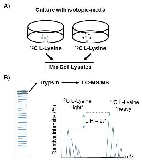
Figure 4: . Stable isotope labeling by amino acids in culture (SILAC) to quantitatively compare the abundance of proteins.

Figure 4:

Stable isotope labeling by amino acids in culture (SILAC) to quantitatively compare the abundance of proteins. A) Two strains (wild type and mutant) are grown with either a 13C or 12C amino acid such as lysine. Following culture, cell lysates are obtained and the proteins combined and separated by gel electrophoresis. B) Protein bands are excised from the gel, digested with trypsin, and analyzed together with LC-MS/MS. The intensity of the peptide ions corresponds to the relative abundance of the parent proteins.

From: The Streptococcal Proteome
Cover of Streptococcus pyogenes
Streptococcus pyogenes : Basic Biology to Clinical Manifestations [Internet].
Ferretti JJ, Stevens DL, Fischetti VA, editors.
© The University of Oklahoma Health Sciences Center.

Except where otherwise noted, this work is licensed under a Creative Commons Attribution-NonCommercial-NoDerivatives 4.0 International License (CC-BY-NC-ND 4.0). To view a copy of this license, visit http://creativecommons.org/licenses/by-nc-nd/4.0/

NCBI Bookshelf. A service of the National Library of Medicine, National Institutes of Health.