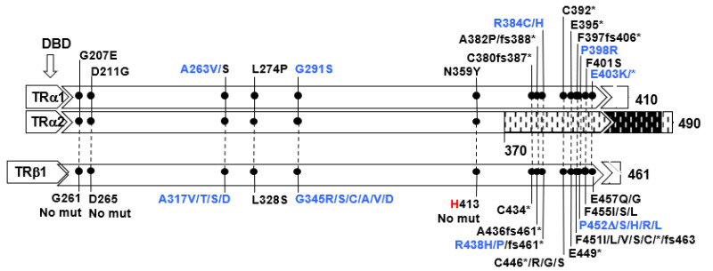
Figure 7. . Mutations in TRα1 and TRα2 and in the corresponding amino acid mutations in TRß1 are aligned according to amino acid sequence homology.

Figure 7.

Mutations in TRα1 and TRα2 and in the corresponding amino acid mutations in TRß1 are aligned according to amino acid sequence homology. The single difference is indicated in red. In blue are mutations occurring in hot spots (CpG or CG-rich regions. The ligand binding domain (LBD) containing the mutations is expanded and the locations of mutation is in scale. The DNA binding domain DLBD) is upstream of the mutations. Sequences from amino acid 370 to 490 of the TRα2 diverge from those of TRα1 due to alternative splicing. Data on THRA gene mutations courtesy of Carla Moran, University of Cambridge, United Kingdom.

From: Impaired Sensitivity to Thyroid Hormone: Defects of Transport, Metabolism, and Action

Cover of Endotext
Endotext [Internet].
Feingold KR, Anawalt B, Blackman MR, et al., editors.
South Dartmouth (MA): MDText.com, Inc.; 2000-.
Copyright © 2000-2024, MDText.com, Inc.

This electronic version has been made freely available under a Creative Commons (CC-BY-NC-ND) license. A copy of the license can be viewed at http://creativecommons.org/licenses/by-nc-nd/2.0/.

NCBI Bookshelf. A service of the National Library of Medicine, National Institutes of Health.