U.S. flag

An official website of the United States government

NCBI Bookshelf. A service of the National Library of Medicine, National Institutes of Health.

StemBook [Internet]. Cambridge (MA): Harvard Stem Cell Institute; 2008-. doi: 10.3824/stembook.1.37.1

Cover of StemBook

StemBook [Internet].

Show details
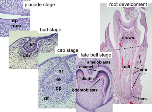
Figure 2. Histology of important stages of tooth development.

Figure 2Histology of important stages of tooth development.

Note that all early development is directed at creating the crown and only then root formation is initiated. Ameloblasts differentiate from the epithelium and odontoblasts from the mesenchyme and they deposit the matrices of enamel and dentin, respectively. Ameloblasts and enamel are missing on the root which is covered by the softer dentin and cementum. Ep, epithelium; mes, mesenchyme; sr, stellate reticulum; dm; dental mesenchyme; dp, dental papilla; df, dental follicle; ek, enamel knot; erm, epithelial cell rests of malassez; hers, hertwig's epithelial root sheath.

From: Tooth organogenesis and regeneration

Copyright: © 2008 Irma Thesleff and Mark Tummers.

This is an open-access article distributed under the terms of the Creative Commons Attribution License, which permits unrestricted use, distribution, and reproduction in any medium, provided the original work is properly cited.


Views

  • Cite this Page
  • PDF version of this page (1.6M)

Related information

Similar articles in PubMed

See reviews...See all...

Recent Activity

Your browsing activity is empty.

Activity recording is turned off.

Turn recording back on

See more...