From: Mitosis

NCBI Bookshelf. A service of the National Library of Medicine, National Institutes of Health.

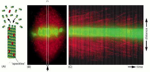
(A) The principle of the method. A very low amount of fluorescent tubulin is injected into living cells so that individual microtubules form with a very small proportion of fluorescent tubulin. Such microtubules have a speckled appearance when viewed by fluorescence microscopy. (B) Fluorescence micrographs of a mitotic spindle in a living newt lung epithelial cell. The chromosomes are stained green, and the tubulin speckles are red. (C) The movement of individual speckles can be readily followed by time-lapse video microscopy. Images of the long, thin, rectangular, boxed region (arrow) in (B) were taken at sequential times and pasted side by side to make a montage of the region over time. Individual speckles can be seen to move toward the poles (representing poleward flux) at a rate of about 0.75 μm/min. (From T.J. Mitchison and E.D. Salmon, Nature Cell Biol. 3:E17–21, 2001.)
From: Mitosis

NCBI Bookshelf. A service of the National Library of Medicine, National Institutes of Health.