NCBI Bookshelf. A service of the National Library of Medicine, National Institutes of Health.
Alberts B, Johnson A, Lewis J, et al. Molecular Biology of the Cell. 4th edition. New York: Garland Science; 2002.

Molecular Biology of the Cell. 4th edition.
Show details
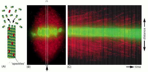
Figure 18-21Visualizing the dynamics of individual microtubules by fluorescence speckle microscopy
(A) The principle of the method. A very low amount of fluorescent tubulin is injected into living cells so that individual microtubules form with a very small proportion of fluorescent tubulin. Such microtubules have a speckled appearance when viewed by fluorescence microscopy. (B) Fluorescence micrographs of a mitotic spindle in a living newt lung epithelial cell. The chromosomes are stained green, and the tubulin speckles are red. (C) The movement of individual speckles can be readily followed by time-lapse video microscopy. Images of the long, thin, rectangular, boxed region (arrow) in (B) were taken at sequential times and pasted side by side to make a montage of the region over time. Individual speckles can be seen to move toward the poles (representing poleward flux) at a rate of about 0.75 μm/min. (From T.J. Mitchison and E.D. Salmon, Nature Cell Biol. 3:E17–21, 2001.)
- Figure 18-21, Visualizing the dynamics of individual microtubules by fluorescenc...Figure 18-21, Visualizing the dynamics of individual microtubules by fluorescence speckle microscopy - Molecular Biology of the Cell
- Methylobacterium cerastii partial 16S rRNA gene, isolate C15Methylobacterium cerastii partial 16S rRNA gene, isolate C15gi|321399889|emb|FR733885.1|Nucleotide
- CR574412 XGC-tailbud-head Xenopus tropicalis cDNA clone THdA023j03 3', mRNA sequ...CR574412 XGC-tailbud-head Xenopus tropicalis cDNA clone THdA023j03 3', mRNA sequencegi|50461838|gnl|dbEST|24659620|emb| 412.1|Nucleotide
- SLC35F6 [Ursus arctos]SLC35F6 [Ursus arctos]Gene ID:113270893Gene
- Cd300c2 CD300C molecule 2 [Mus musculus]Cd300c2 CD300C molecule 2 [Mus musculus]Gene ID:140497Gene
Your browsing activity is empty.
Activity recording is turned off.
See more...