From: Chapter 9, Benign Childhood Focal Seizures and Related Epileptic Syndromes

NCBI Bookshelf. A service of the National Library of Medicine, National Institutes of Health.

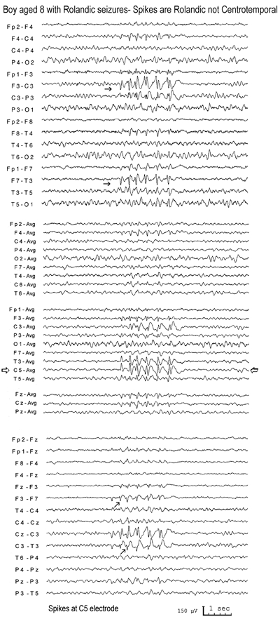
Centrotemporal spikes are mainly Rolandic not temporal spikes.1;35 Facing page
Top, middle and bottom: The same EEG sample is shown in 3 different montages.
This is from an 8-year-old boy referred for an EEG because of “recent GTCS and a 2-year history of unilateral facial spasms. Previously, the EEG and CT brain scan were normal. No medication. Focal seizures with secondarily generalised convulsions?”
The EEG showed frequent clusters of repetitive centrotemporal spikes on the left. Because the spikes appeared to be of higher amplitude in the temporal electrode (T3) (black arrows), the technologist rightly applied additional electrodes at C5 and C6 (Rolandic localisation). This showed that the spike is of higher amplitude in the left Rolandic region (C5) (open arrows). Another EEG 16 months later, showed a few small spikes in the right frontal and central midline electrodes.
From: Chapter 9, Benign Childhood Focal Seizures and Related Epileptic Syndromes

NCBI Bookshelf. A service of the National Library of Medicine, National Institutes of Health.