

NCBI Bookshelf. A service of the National Library of Medicine, National Institutes of Health.
Coffin JM, Hughes SH, Varmus HE, editors. Retroviruses. Cold Spring Harbor (NY): Cold Spring Harbor Laboratory Press; 1997.
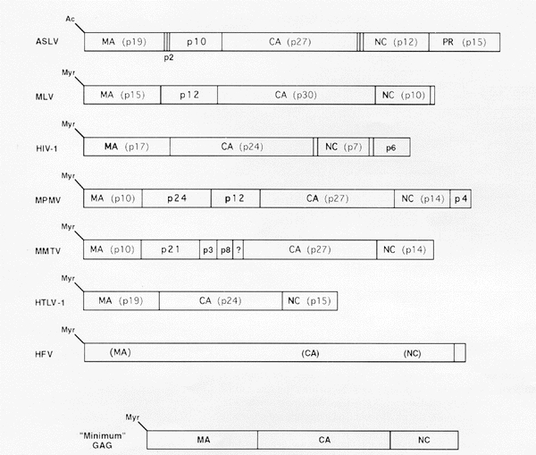
Figure 7
Organization of Gag proteins. Schematic representations of Gag proteins are drawn for examples from each retroviral genus. Vertical solid lines mark cleavage sites for the viral protease. The sequences representing the mature proteins MA, CA, NC, and PR are indicated, along with the older naming of these proteins based on their approximate molecular weight. (Ac) Acetylation at the amino terminus; (Myr) myristylation at the amino terminus.
- Figure 7, [Organization of Gag proteins. Schematic...]. - RetrovirusesFigure 7, [Organization of Gag proteins. Schematic...]. - Retroviruses
Your browsing activity is empty.
Activity recording is turned off.
See more...