All rights reserved. Publications of the World Health Organization are available on the WHO web site (www.who.int) or can be purchased from WHO Press, World Health Organization, 20 Avenue Appia, 1211 Geneva 27, Switzerland (tel.: +41 22 791 3264; fax: +41 22 791 4857; e-mail: tni.ohw@sredrokoob). Requests for permission to reproduce or translate WHO publications – whether for sale or for non-commercial distribution – should be addressed to WHO Press through the WHO web site (www.who.int/about/licensing/copyright_form/en/index.html).
NCBI Bookshelf. A service of the National Library of Medicine, National Institutes of Health.
Pocket Book of Hospital Care for Children: Guidelines for the Management of Common Childhood Illnesses. 2nd edition. Geneva: World Health Organization; 2013.
Pocket Book of Hospital Care for Children: Guidelines for the Management of Common Childhood Illnesses. 2nd edition.
Show detailsCough and difficulty in breathing are common problems in young children. The causes range from a mild, self-limited illness to severe, life-threatening disease. This chapter provides guidelines for managing the most important conditions that cause cough, difficulty in breathing or both in children aged 2 months to 5 years. The differential diagnosis of these conditions is described in Chapter 2. Management of these problems in infants < 2 months of age is described in Chapter 3 and management in severely malnourished children in Chapter 7.
Most episodes of cough are due to the common cold, each child having several episodes a year. The commonest severe illness and cause of death that presents with cough or difficult breathing is pneumonia, which should be considered first in any differential diagnosis (Table 6).
Table 6Differential diagnosis in a child presenting with cough or difficulty in breathing
| Diagnosis | In favour |
|---|---|
| Pneumonia |
|
| Effusion or empyema |
|
| Asthma or wheeze |
|
| Bronchiolitis |
|
| Malaria |
|
| Severe anaemia |
|
| Cardiac failure |
|
| Congenital heart disease (cyanotic) |
|
| Congenital heart disease (acyanotic) |
|
| Tuberculosis |
|
| Pertussis |
|
| Foreign body |
|
| Pneumothorax |
|
| Pneumocystis pneumonia |
|
| Croup |
|
| Diphtheria |
|
4.1. Child presenting with cough
History
Pay particular attention to:
- cough
- –
duration in days
- –
paroxysms with whoops or vomiting or central cyanosis
- exposure to someone with TB (or chronic cough) in the family
- history of choking or sudden onset of symptoms
- known or possible HIV infection
- vacci nation history: BCG; diphtheria, pertussis, tetanus (DPT); measles; Haemophilus influenzae type b and pneumococcus
- personal or family history of asthma.
Examination
The symptoms and signs listed below are a guide for the clinician to reach a diagnosis. Not all children will show every symptom or sign.
General
- central cyanosis
- apnoea, gasping, grunting, nasal flaring, audible wheeze, stridor
- head nodding (a movement of the head synchronous with inspiration indicating severe respiratory distress)
- tachycardia
- severe palmar pallor
Chest
- respiratory rate (count during 1 min when the child is calm)
fast breathing: < 2 months, ≥ 60 breaths 2–11 months, ≥ 50 breaths 1–5 years, ≥ 40 breaths - lower chest wall indrawing
- hyperinflated chest
- apex beat displaced or trachea shifted from midline
- raised jugular venous pressure
- on auscultation, coarse crackles, no air entry or bronchial breath sounds or wheeze
- abnormal heart rhythm on auscultation
- percussion signs of pleural effusion (stony dullness) or pneumothorax (hyper-resonance)
Note: Lower chest wall indrawing is when the lower chest wall goes in when the child breathes in; if only the soft tissue between the ribs or above the clavicle goes in when the child breathes, this is not lower chest wall indrawing.
Abdomen
- abdominal masses (e.g. lymphadenopathy)
- enlarged liver and spleen
Investigations
- pulse oximetry to detect hypoxia and as a guide to when to start or stop oxygen therapy
- full blood count
- chest X-ray only for children with severe pneumonia or pneumonia that does not respond to treatment or complications or unclear diagnosis or associated with HIV.
4.2. Pneumonia
Pneumonia is caused by viruses or bacteria. It is usually not possible to determine the specific cause of pneumonia by clinical features or chest X-ray appearance. Pneumonia is classified as severe or non-severe on the basis of clinical features, the management being based on the classification. Antibiotic therapy should be given in most cases of pneumonia and severe pneumonia. Severe pneumonia may require additional supportive care, such as oxygen, to be given in hospital.
4.2.1. Severe pneumonia
Diagnosis
Cough or difficulty in breathing, plus at least one of the following:
- central cyanosis or oxygen saturation < 90% on pulse oximetry
- severe respiratory distress (e.g. grunting, very severe chest indrawing)
- signs of pneumonia with a general danger sign:
- –
inability to breastfeed or drink,
- –
lethargy or unconscious,
- –
convulsions.
- In addition, some or all of the other signs of pneumonia may be present, such as:
- –
signs of pneumonia
fast breathing: age 2–11 months, ≥ 50/min age 1–5 years, ≥ 40/min
- –
chest indrawing: lower chest wall indrawing (i.e. lower chest wall goes in when the child breathes in)
- –
chest auscultation signs:
- decreased breath sounds
- bronchial breath sounds
- crackles
- abnormal vocal resonance (decreased over a pleural effusion or empyema, increased over lobar consolidation)
- pleural rub
Table 7Classification of the severity of pneumonia
| Sign or symptom | Classification | Treatment |
|---|---|---|
Cough or difficulty in breathing with:
| Severe pneumonia |
|
| Pneumonia |
|
| No pneumonia: cough or cold |
|
Investigations
- Measure oxygen saturation with pulse oximetry in all children suspected of having pneumonia.
- If possible, obtain a chest X-ray to identify pleural effusion, empyema, pneumothorax, pneumatocoele, interstitial pneumonia or pericardial effusion.
Treatment
- ►
Admit the child to hospital.
Oxygen therapy
Ensure continuous oxygen supply, either as cylinders or oxygen concentrator, at all times.
- ►
Give oxygen to all children with oxygen saturation < 90%
- ►
Use nasal prongs as the preferred method of oxygen delivery to young infants; if not available, a nasal or nasopharyngeal catheter may be used. The different methods of oxygen administration and diagrams showing their use are given in section 10.7.
- ►
Use a pulse oximetry to guide oxygen therapy (to keep oxygen saturation > 90%). If a pulse oximeter is not available, continue oxygen until the signs of hypoxia (such as inability to breastfeed or breathing rate ≥ 70/min) are no longer present.
- ►
Remove oxygen for a trial period each day for stable children while continuing to use a pulse oximeter to determine oxygen saturation. Discontinue oxygen if the saturation remains stable at > 90% (at least 15 min on room air).
Nurses should check every 3 h that the nasal prongs are not blocked with mucus and are in the correct place and that all connections are secure.
Antibiotic therapy
- ►
Give intravenous ampicillin (or benzylpenicillin) and gentamicin.
- –
Ampicillin 50 mg/kg or benzylpenicillin 50 000 U/kg IM or IV every 6 h for at least 5 days
- –
Gentamicin 7.5 mg/kg IM or IV once a day for at least 5 days.
- ►
If the child does not show signs of improvement within 48 h and staphylococcal pneumonia is suspected, switch to gentamicin 7.5 mg/kg IM or IV once a day and cloxacillin 50 mg/kg IM or IV every 6 h.
- ►
Use ceftriaxone (80 mg/kg IM or IV once daily) in cases of failure of first-line treatment.
Supportive care
- ►
Remove by gentle suction any thick secretions at the entrance to the nasal passages or throat, which the child cannot clear.
- ►
If the child has fever (≥ 39 °C or ≥ 102.2 °F) which appears to be causing distress, give paracetamol.
- ►
If wheeze is present, give a rapid-acting bronchodilator (see below), and start steroids when appropriate.
- ►
Ensure that the child receives daily maintenance fluids appropriate for his or her age (see section 10.2, but avoid over-hydration.
- –
Encourage breastfeeding and oral fluids.
- –
If the child cannot drink, insert a nasogastric tube and give maintenance fluids in frequent small amounts. If the child is taking fluids adequately by mouth, do not use a nasogastric tube as it increases the risk for aspiration pneumonia and obstructs part of the nasal airway. If oxygen is given by nasal catheter at the same time as nasogastric fluids, pass both tubes through the same nostril.
- ►
Encourage the child to eat as soon as food can be taken.
Monitoring
The child should be checked by a nurse at least every 3 h and by a doctor at least twice a day. In the absence of complications, within 2 days there should be signs of improvement (breathing slower, less indrawing of the lower chest wall, less fever, improved ability to eat and drink, better oxygen saturation).
Other alternative diagnosis and treatment
- If the child has not improved after 2 days or if the child's condition has worsened, look for complications (see section 4.3) or alternative diagnoses. If possible, obtain a chest X-ray. The commonest other possible diagnoses are:
Staphylococcal pneumonia. This is suggested if there is rapid clinical deterioration despite treatment, by a pneumatocoele or pneumothorax with effusion on chest X-ray, numerous Gram-positive cocci in a smear of sputum or heavy growth of S. aureus in cultured sputum or empyema fluid. The presence of septic skin pustules supports the diagnosis.
- ►
Treat with cloxacillin (50 mg/kg IM or IV every 6 h) and gentamicin (7.5 mg/kg IM or IV once a day). When the child improves (after at least 7 days of IV or IM antibiotics), continue cloxacillin orally four times a day for a total course of 3 weeks. Note that cloxacillin can be replaced by another anti-staphylococcal antibiotic, such as oxacillin, flucloxacillin or dicloxacillin.
Tuberculosis. A child with persistent cough and fever for more than 2 weeks and signs of pneumonia after adequate antibiotic treatment should be evaluated for TB. If another cause of the fever cannot be found, TB should be considered, particularly in malnourished children. Further investigations and treatment for TB, following national guidelines, may be initiated and response to anti-TB treatment evaluated (see section 4.7.2). The HIV status of all children suspected of having TB should be confirmed if not known.
HIV infection or exposure to HIV. Some aspects of antibiotic treatment are different for children who are HIV positive or in whom HIV infection is suspected. Although pneumonia in many of these children has the same etiology as that in children without HIV, Pneumocystis pneumonia (PCP), often at the age of 4–6 months (see section 8.4) is an important cause to be suspected and treated.
- ►
Treat as for severe pneumonia above; give ampicillin plus gentamicin IM or IV for 10 days.
- ►
If the child does not improve within 48 h, switch to ceftriaxone at 80 mg/kg IV once daily over 30 min. If ceftriaxone is not available, give gentamicin plus cloxacillin, as above.
- ►
For children < 12 months, also give high-dose co-trimoxazole (8 mg/kg trimethoprim and 40 mg/kg sulfamethoxazole IV every 8 h or orally three times a day) for 3 weeks. For a child aged 12–59 months, give this treatment only if there are clinical signs of PCP (such as chest X-ray findings of interstitial pneumonia).
- ►
For further management of the child, including PCP prophylaxis, see Chapter 8).
Discharge
Children with severe pneumonia can be discharged when:
- Respiratory distress has resolved.
- There is no hypoxaemia (oxygen saturation, > 90%).
- They are feeding well.
- They are able to take oral medication or have completed a course of parenteral antibiotics.
- The parents understand the signs of pneumonia, risk factors and when to return.

Normal chest X-ray
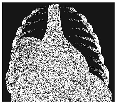
Lobar pneumonia of the right lower zone indicated by a consolidation (X-ray)

Staphylococcal pneumonia
Typical features include pneumatocoeles (right), and an abscess with an air-fluid level (left) (X-ray).

Pneumothorax
The right lung (left side on image) is collapsed towards the hilus, leaving a transparent margin without lung structure. In contrast, the right side (normal) demonstrates markings extending to the periphery (X-ray).

Hyperinflated chest
Features are an increased transverse diameter, ribs running more horizontally, a small contour of the heart, and flattened diaphragm (X-ray).
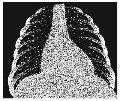
Appearance of miliary tuberculosis: widespread small patchy infiltrates throughout both lungs: “snow storm appearance” (X-ray)
Follow-up
Children with severe pneumonia may cough for several weeks. As they have been very sick, their nutrition is often poor. Give the vaccinations that are due, and arrange follow-up 2 weeks after discharge, if possible, to check the child's nutrition. Also address risk factors such as malnutrition, indoor air pollution and parental smoking.
4.2.2. Pneumonia
Diagnosis
Cough or difficult breathing plus at least one of the following signs:
fast breathing: age 2–11 months, ≥ 50/min age 1–5 years, ≥ 40/min - lower chest wall indrawing
In addition, either crackles or pleural rub may be present on chest auscultation.
Check that there are no signs of severe pneumonia, such as:
- –
oxygen saturation < 90% on pulse oximetry or central cyanosis
- –
severe respiratory distress (e.g. grunting, very severe chest indrawing)
- –
inability to breastfeed or drink or vomiting everything
- –
convulsions, lethargy or reduced level of consciousness
- –
auscultatory findings of decreased or bronchial breath sounds or signs of pleural effusion or empyema.
Treatment
- ►
Treat child as outpatient.
- ►
Advise carers to give normal fluid requirements plus extra breast milk or fluids if there is a fever. Small frequent drinks are more likely to be taken and less likely to be vomited
Antibiotic therapy
- ►
Give the first dose at the clinic and teach the mother how to give the other doses at home.
- ►
Give oral amoxicillin:
- –
In settings with high HIV infection rate, give oral amoxicillin at least 40 mg/kg per dose twice a day for 5 days.
- –
In areas with low HIV prevalence, give amoxicillin at least 40 mg/kg per dose twice a day for 3 days.
- ►
Avoid unnecessary harmful medications such as remedies containing atropine, codeine derivatives or alcohol.

Lower chest wall indrawing: with inspiration, the lower chest wall moves in
Follow-up
Encourage the mother to feed the child. Advise her to bring the child back after 3 days, or earlier if the child becomes sicker or is unable to drink or breastfeed. When the child returns, check:
- Whether the breathing has improved (slower), there is no chest indrawing, less fever, and the child is eating better; complete the antibiotic treatment.
- If the breathing rate and/or chest indrawing or fever and/or eating have not improved, exclude a wheeze. If no wheeze, admit to hospital for investigations to exclude complications or alternative diagnosis.
- If signs of severe pneumonia are present, admit the child to hospital and treat as above.
- Address risk factors such as malnutrition, indoor air pollution and parental smoking.
Pneumonia in children with HIV infection
- ►
Admit to hospital and manage as severe pneumonia (see section 4.2.1).
- ►
For further management of these children, including PCP prophylaxis (see Chapter 8).
4.3. Complications of pneumonia
Septicaemia is the most common pneumonia complication and occurs when the bacteria causing pneumonia spreads into the bloodstream (see section 6.5). The spread of bacteria can lead to septic shock or metastatic secondary infections like meningitis especially in infants, peritonitis, and endocarditis especially in patients with vulvar heart disease or septic arthritis. Other common complication include pleural effusion, empyema and lung abscess.
4.3.1. Pleural effusion and empyema
Diagnosis
A child with pneumonia may develop pleural effusion or empyema.
- On examination, the chest is dull to percussion, and breath sounds are reduced or absent over the affected area.
- A pleural rub may be heard at an early stage before the effusion is fully developed.
- A chest X-ray shows fluid on one or both sides of the chest.
- When empyema is present, fever persists despite antibiotic therapy, and the pleural fluid is cloudy or frankly purulent.
Treatment
Drainage
- ►
Pleural effusions should be drained, unless they are very small. If effusions are present on both sides of the chest, drain both. It may be necessary to repeat drainage two or three times if fluid returns. See Annex A1.5, for guidelines on chest drainage.
Subsequent management depends on the character of the fluid obtained. When possible, pleural fluid should be analysed for protein and glucose content, cell count and differential count, and examined after Gram and Ziehl-Neelsen staining and bacterial and Mycobacterium tuberculosis culture.
Antibiotic therapy
- ►
Give ampicillin or cloxacillin or flucloxacillin (50 mg/kg IM or IV every 6 h) and gentamicin (7.5 mg/kg IM or IV once a day). When the child improves (after at least 7 days of IV or IM antibiotics), continue cloxacillin orally four times a day for a total course of 3 weeks.
Note: Cloxacillin is preferable if staphylococcal infection is suspected; it can be replaced by another anti-staphylococcal antibiotic such as oxacillin, flucloxacillin or dicloxacillin. Infection with S. aureus is more likely if pneumatocoeles are also present.
Failure to improve
If fever and other signs of illness continue, despite adequate chest drainage and antimicrobial therapy, test for HIV infection and assess for possible TB.
- ►
A trial of anti-TB therapy may be required (see section 4.7.2).
4.3.2. Lung abscess
A lung abscess is a circumscribed, thick-walled cavity in the lung that contains purulent material resulting from suppuration and necrosis of the involved lung parenchyma. It frequently develops in an unresolved area of pneumonia. This could be a result of pulmonary aspiration, diminished clearance mechanisms, embolic phenomena, or haematogenous spread.
Diagnosis
Common signs and symptoms:
- Fever
- Pleuritic chest pain
- Sputum production or haemoptysis
- Weight loss
- On examination: reduced chest movement, decreased breath sounds, dullness to percussion, crackles, and bronchial breathing.
- Chest X-ray: solitary, thick-walled cavity in the lung with or without air fluid level.
- Ultrasonography and CT scan: to localize the lesion and guide drainage or needle aspiration.
Treatment
The choice of antibiotic is usually empirical and is based on the underlying condition of the patient and the presumed etiological agent.
- ►
Give ampicillin or cloxacillin or flucloxacillin (50 mg/kg IM or IV every 6 h) and gentamicin (7.5 mg/kg IM or IV once a day). Continue treatment as in empyema (see section 4.3.1) for up to 3 weeks.
- ►
Surgical management is considered in cases of large lung abscess especially when associated with haemoptysis or clinical deterioration despite appropriate antibiotic therapy. Drainage is usually through percutaneous tube drainage or ultrasound guided needle aspiration.
4.3.3. Pneumothorax
Pneumothorax is usually secondary to an accumulation of air in the pleural spaces from alveolar rupture or from infection with gas-producing microorganisms.
Diagnosis
- Signs and symptoms may vary according to the extent of lung collapse, degree of intrapleural pressure, and rapidity of onset.
- On examination: chest bulging on the affected side if one side is involved, shift of cardiac impulse away from the site of the pneumothorax, decreased breath sounds on the affected side, grunting, severe respiratory distress and cyanosis may occur late in the progression of the complication.
- Differential diagnosis include lung cyst, lobar emphysema, bullae, diaphragmatic hernia
- Chest X-ray is crucial in the confirmation of diagnosis.
Treatment
- ►
Insert needle for urgent decompression, before insertion of an intercostal chest drain.
See Annex A1.5, for guidelines on chest drainage.
4.4. Cough or cold
These are common, self-limited viral infections that require only supportive care. Antibiotics should not be given. Wheeze or stridor may occur in some children, especially infants. Most episodes end within 14 days. Cough lasting 14 days or more may be caused by TB, asthma, pertussis or symptomatic HIV infection (see Chapter 8).
Diagnosis
Common features:
- cough
- nasal discharge
- mouth breathing
- fever
The following are absent:
- –
general danger signs.
- –
signs of severe pneumonia or pneumonia
- –
stridor when the child is calm
Wheezing may occur in young children (see below).
Treatment
- ►
Treat the child as an outpatient.
- ►
Soothe the throat and relieve the cough with a safe remedy, such as a warm, sweet drink.
- ►
Relieve high fever (≥ 39 °C or ≥ 102.2 °F) with paracetamol if the fever is causing distress to the child.
- ►
Clear secretions from the child's nose before feeds with a cloth soaked in water that has been twisted to form a pointed wick.
Give normal fluid requirements plus extra breast milk or fluids if there is fever. Small frequent drinks are more likely to be taken and less likely to be vomited.
- ►
Do not give any of the following:
- –
an antibiotic (they are not effective and do not prevent pneumonia)
- –
remedies containing atropine, codeine or codeine derivatives, or alcohol (these may be harmful) or mucolytics
- –
medicated nose drops.
Follow-up
Advise the mother to:
- feed the child
- watch for fast or difficult breathing and return if either develops
- return if the child becomes sicker or is unable to drink or breastfeed.
4.5. Conditions presenting with wheeze
Wheeze is a high-pitched whistling sound on expiration. It is caused by spasmodic narrowing of the distal airway. To hear a wheeze, even in mild cases, place your ear next to the child's mouth and listen to the breathing while the child is calm, or use a stethoscope.
In the first 2 years of life, wheezing is most commonly caused by acute viral respiratory infections such as bronchiolitis or coughs and colds. After 2 years of age, most wheezing is due to asthma (Table 8). Some children with pneumonia present with wheeze. It is important always to consider treatment for pneumonia, particularly in the first 2 years of life. Children with wheeze but no fever, chest indrawing or danger signs are unlikely to have pneumonia and should therefore not be given antibiotics.
Table 8Differential diagnosis in a child presenting with wheeze
| Diagnosis | In favour |
|---|---|
| Asthma |
|
| Bronchiolitis |
|
| Wheeze associated with cough or cold |
|
| Foreign body |
|
| Pneumonia |
|
History
- previous episodes of wheeze
- night-time or early morning shortness of breath, cough or wheeze
- response to bronchodilators
- asthma diagnosis or long-term treatment for asthma
- family history of allergy or asthma
Examination
- wheezing on expiration
- prolonged expiration
- resonant percussion note
- hyperinflated chest
- rhonchi on auscultation
- shortness of breath at rest or on exertion
- lower chest wall indrawing if severe.
Response to rapid-acting bronchodilator
- ►
If the cause of the wheeze is not clear or if the child has fast breathing or chest indrawing in addition to wheeze, give a rapid-acting bronchodilator and assess after 15 min. The response to a rapid-acting bronchodilator helps to determine the underlying diagnosis and treatment.
- ►
Give the rapid-acting bronchodilator by one of the following methods:
- –
nebulized salbutamol
- –
salbutamol by a metered dose inhaler with spacer device
- –
if neither of the above methods is available, give a subcutaneous injection of adrenaline.
For details of administering the above, see below.
- ■
Assess the response after 15 min. Signs of improvement are:
- –
less respiratory distress (easier breathing)
- –
less lower chest wall indrawing
- –
improved air entry.
- ►
Children who still have signs of hypoxia (central cyanosis, low oxygen saturation ≤ 90%, unable to drink due to respiratory distress, severe lower chest wall indrawing) or have fast breathing should be given a second dose of bronchodilator and admitted to hospital for further treatment.
4.5.1. Bronchiolitis
Bronchiolitis is a lower respiratory viral infection, which is typically most severe in young infants, occurs in annual epidemics and is characterized by airways obstruction and wheezing. It is most commonly caused by respiratory syncytial virus. Secondary bacterial infection may occur. The management of bronchiolitis associated with fast breathing or other sign of respiratory distress is therefore similar to that of pneumonia. Episodes of wheeze may occur for months after an attack of bronchiolitis, but will eventually stop.
Diagnosis
Typical features of bronchiolitis, on examination, include:
- wheezing that is not relieved by up to three doses of a rapid-acting bronchodilator
- hyperinflation of the chest, with increased resonance to percussion
- lower chest wall indrawing
- fine crackles and wheeze on auscultation of the chest
- difficulty in feeding, breastfeeding or drinking owing to respiratory distress
- nasal discharge, which can cause severe nasal obstruction.
Treatment
Most children can be treated at home, but those with the following signs of severe pneumonia (see section 4.2.1) should be treated in hospital:
- oxygen saturation < 90% or central cyanosis.
- apnoea or history of apnoea
- inability to breastfeed or drink, or vomiting everything
- convulsions, lethargy or unconsciousness
- gasping and grunting (especially in young infants).
Oxygen
- ►
Give oxygen to all children with severe respiratory distress or oxygen saturation ≤ 90% (see section 4.2.1). The recommended method for delivering oxygen is by nasal prongs or a nasal catheter.
- ►
The nurse should check, every 3 h, that the prongs are in the correct position and not blocked with mucus, and that all connections are secure.
Antibiotic treatment
- ►
If the infant is treated at home, give amoxicillin (40 mg/kg twice a day) orally for 5 days only if the child has signs of pneumonia (fast breathing and lower chest wall indrawing).
- ►
If there are signs of severe pneumonia, give ampicillin at 50 mg/kg or benzylpenicillin at 50 000 U/kg IM or IV every 6 h for at least 5 days and gentamicin 7.5 mg/kg IM or IV once a day for at least 5 days.
Supportive care
- ►
If the child has fever (≥ 39 °C or ≥ 102.2 °F) that appears to be causing distress, give paracetamol.
- ►
Ensure that the hospitalized child receives daily maintenance fluids appropriate for age (see section 10.2), but avoid overhydration. Encourage breastfeeding and oral fluids.
- ►
Encourage the child to eat as soon as food can be taken. Nasogastric feeding should be considered in any patient who is unable to maintain oral intake or hydration (expressed breast milk is the best).
- ►
Gentle nasal suction should be used to clear secretions in infants where nasal blockage appears to be causing respiratory distress.
Monitoring
A hospitalized child should be assessed by a nurse every 6 h (or every 3 h if there are signs of very severe illness) and by a doctor at least once a day. Monitor oxygen therapy as described in section 10.7. Watch for signs of respiratory failure, i.e. increasing hypoxia and respiratory distress leading to exhaustion.
Complications
If the child fails to respond to oxygen therapy or the child's condition worsens suddenly, obtain a chest X-ray to look for evidence of pneumothorax.
Tension pneumothorax associated with severe respiratory distress and shift of the heart requires immediate relief by placing a needle to allow the air that is under pressure to escape (needle thoracocentesis). Following this, a continuous air exit should be assured by inserting a chest tube with an underwater seal until the air leak closes spontaneously and the lung expands (see Annex A1.5).
If respiratory failure develops, continuous positive airway pressure may be helpful.
Infection control
Bronchiolitis is very infectious and dangerous to other young children in hospital with other conditions. The following strategies may reduce cross-infection:
- hand-washing by personnel between patients
- ideally isolate the child, but maintain close observation
- during epidemics, restrict visits to children by parents and siblings with symptoms of upper respiratory tract infection.
Discharge
An infant with bronchiolitis can be discharged when respiratory distress and hypoxaemia have resolved, when there is no apnoea and the infant is feeding well. Infants are at risk for recurrent bronchiolitis if they live in families where adults smoke or if they are not breastfed. So, advise the parents against smoking.
Follow-up
Infants with bronchiolitis may have cough and wheeze for up to 3 weeks. As long as they are well with no respiratory distress, fever or apnoea and are feeding well they do not need antibiotics.
4.5.2. Asthma
Asthma is a chronic inflammatory condition with reversible airways obstruction. It is characterized by recurrent episodes of wheezing, often with cough, which respond to treatment with bronchodilators and anti-inflammatory drugs. Antibiotics should be given only when there are signs of pneumonia.
Diagnosis
History of recurrent episodes of wheezing, often with cough, difficulty in breathing and tightness in the chest, particularly if these are frequent and recurrent or are worse at night and in the early morning. Findings on examination may include:
- rapid or increasing respiratory rate
- hyperinflation of the chest
- hypoxia (oxygen saturation ≤ 90%)
- lower chest wall indrawing
- use of accessory muscles for respiration (best noted by feeling the neck muscles)
- prolonged expiration with audible wheeze
- reduced or no air intake when obstruction is life-threatening
- absence of fever
- good response to treatment with a bronchodilator.
If the diagnosis is uncertain, give a dose of a rapid-acting bronchodilator (see salbutamol). A child with asthma will often improve rapidly with such treatment, showing signs such as slower respiratory rate, less chest wall indrawing and less respiratory distress. A child with severe asthma may require several doses in quick succession before a response is seen (see below).
Treatment
- ►
A child with a first episode of wheezing and no respiratory distress can usually be managed at home with supportive care. A bronchodilator is not necessary.
- ►
If the child is in respiratory distress (acute severe asthma) or has recurrent wheezing, give salbutamol by metered-dose inhaler and spacer device or, if not available, by nebulizer (see below for details). If salbutamol is not available, give subcutaneous adrenaline.
- ►
Reassess the child after 15 min to determine subsequent treatment:
- –
If respiratory distress has resolved, and the child does not have fast breathing, advise the mother on home care with inhaled salbutamol from a metered dose inhaler and spacer device (which can be made locally from plastic bottles).
- –
If respiratory distress persists, admit to hospital and treat with oxygen, rapid-acting bronchodilators and other drugs, as described below.
Severe life-threatening asthma
- ►
If the child has life-threatening acute asthma, is in severe respiratory distress with central cyanosis or reduced oxygen saturation ≤ 90%, has poor air entry (silent chest), is unable to drink or speak or is exhausted and confused, admit to hospital and treat with oxygen, rapid-acting bronchodilators and other drugs, as described below.
- ►
In children admitted to hospital, promptly give oxygen, a rapid-acting bronchodilator and a first dose of steroids.
Oxygen
- ►
Give oxygen to keep oxygen saturation > 95% in all children with asthma who are cyanosed (oxygen saturation ≤ 90%) or whose difficulty in breathing interferes with talking, eating or breastfeeding.
Rapid-acting bronchodilators
- ►
Give the child a rapid-acting bronchodilator, such as nebulized salbutamol or salbutamol by metered-dose inhaler with a spacer device. If salbutamol is not available, give subcutaneous adrenaline, as described below.
Nebulized salbutamol
The driving source for the nebulizer must deliver at least 6–9 litres/min. Recommended methods are an air compressor, ultrasonic nebulizer or oxygen cylinder, but in severe or life-threatening asthma oxygen must be used. If these are not available, use an inhaler and spacer. An easy-to-operate foot pump may be used but is less effective.
- ►
Put the dose of the bronchodilator solution in the nebulizer compartment, add 2–4 ml of sterile saline and nebulize the child until the liquid is almost all used up. The dose of salbutamol is 2.5 mg (i.e. 0.5 ml of the 5 mg/ml nebulizer solution).
- ►
If the response to treatment is poor, give salbutamol more frequently.
- ►
In severe or life-threatening asthma, when a child cannot speak, is hypoxic or tiring with lowered consciousness, give continuous back-to-back nebulizers until the child improves, while setting up an IV cannula. As asthma improves, a nebulizer can be given every 4 h and then every 6–8 h.
Giving salbutamol by metered-dose inhaler with a spacer device
Spacer devices with a volume of 750 ml are commercially available.
- ►
Introduce two puffs (200 μg) into the spacer chamber. Then, place the child's mouth over the opening in the spacer and allow normal breathing for three to five breaths. This can be repeated in rapid succession until six puffs of the drug have been given to a child < 5 years, 12 puffs for > 5 years of age. After 6 or 12 puffs, depending on age, assess the response and repeat regularly until the child's condition improves. In severe cases, 6 or 12 puffs can be given several times an hour for a short period.
Some infants and young children cooperate better when a face mask is attached to the spacer instead of the mouthpiece.
If commercial devices are not available, a spacer device can be made from a plastic cup or a 1-litre plastic bottle. These deliver three to four puffs of salbutamol, and the child should breathe from the device for up to 30 s.
Subcutaneous adrenaline
- ►
If the above two methods of delivering salbutamol are not available, give a subcutaneous injection of adrenaline at 0.01 ml/kg of 1:1000 solution (up to a maximum of 0.3 ml), measured accurately with a 1-ml syringe (for injection technique, see annex 1). If there is no improvement after 15 min, repeat the dose once.
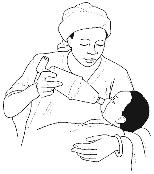
Use of spacer device and face mask to give bronchodilator treatment
A spacer can be made locally from a plastic soft-drink bottle.
Steroids
- ►
If a child has a severe or life-threatening acute attack of wheezing (asthma), give oral prednisolone, 1 mg/kg, for 3–5 days (maximum, 60 mg) or 20 mg for children aged 2–5 years. If the child remains very sick, continue the treatment until improvement is seen.
Repeat the dose of prednisolone for children who vomit, and consider IV steroids if the child is unable to retain orally ingested medication. Treatment for up to 3 days is usually sufficient, but the duration should be tailored to bring about recovery. Tapering of short courses (7–14 days) of steroids is not necessary. IV hydrocortisone (4 mg/kg repeated every 4 h) provides no benefit and should be considered only for children who are unable to retain oral medication.
Magnesium sulfate
Intravenous magnesium sulfate may provide additional benefit in children with severe asthma treated with bronchodilators and corticosteroids. Magnesium sulfate has a better safety profile in the management of acute severe asthma than aminophylline. As it is more widely available, it can be used in children who are not responsive to the medications described above.
- ►
Give 50% magnesium sulfate as a bolus of 0.1 ml/kg (50 mg/kg) IV over 20 min.
Aminophylline
Aminophylline is not recommended in children with mild-to-moderate acute asthma. It is reserved for children who do not improve after several doses of a rapid-acting bronchodilator given at short intervals plus oral prednisolone. If indicated in these circumstances:
- ►
Admit the child ideally to a high-care or intensive-care unit, if available, for continuous monitoring.
- ►
Weigh the child carefully and then give IV aminophylline at an initial loading dose of 5–6 mg/kg (up to a maximum of 300 mg) over at least 20 min but preferably over 1 h, followed by a maintenance dose of 5 mg/kg every 6 h.
IV aminophylline can be dangerous at an overdose or when given too rapidly.
- Omit the initial dose if the child has already received any form of aminophylline or caffeine in the previous 24 h.
- Stop giving it immediately if the child starts to vomit, has a pulse rate > 180/min, develops a headache or has a convulsion.
Oral bronchodilators
Use of oral salbutamol (in syrup or tablets) is not recommended in the treatment of severe or persistent wheeze. It should be used only when inhaled salbutamol is not available for a child who has improved sufficiently to be discharged home.
Dosage:
- –
Age 1 month to 2 years: 100 μg/kg (maximum, 2 mg) up to four times daily
- –
Age 2–6 years: 1–2 mg up to four times daily
Antibiotics
- ►
Antibiotics should not be given routinely for asthma or to a child with asthma who has fast breathing without fever. Antimicrobial treatment is indicated, however, when there is persistent fever and other signs of pneumonia (see section 4.2).
Supportive care
- ►
Ensure that the child receives daily maintenance fluids appropriate for his or her age. Encourage breastfeeding and oral fluids. Encourage adequate complementary feeding for the young child, as soon as food can be taken.
Monitoring
A hospitalized child should be assessed by a nurse every 3 h or every 6 h as the child shows improvement (i.e. slower breathing rate, less lower chest wall indrawing and less respiratory distress) and by a doctor at least once a day. Record the respiratory rate, and watch especially for signs of respiratory failure – increasing hypoxia and respiratory distress leading to exhaustion. Monitor oxygen therapy as described in section 10.7.
Complications
- ►
If the child fails to respond to the above therapy, or the child's condition worsens suddenly, obtain a chest X-ray to look for evidence of pneumothorax. Be very careful in making this diagnosis as the hyperinflation in asthma can mimic a pneumothorax on a chest X-ray. Treat as described in section 4.3.3.
Follow-up care
Asthma is a chronic and recurrent condition.
- ►
Once the child has improved sufficiently to be discharged home, inhaled salbutamol through a metered dose inhaler should be prescribed with a suitable (not necessarily commercial) spacer and the mother instructed on how to use it.
- ►
A long-term treatment plan should be made on the basis of the frequency and severity of symptoms. This may include intermittent or regular treatment with bronchodilators, regular treatment with inhaled steroids or intermittent courses of oral steroids. Up-to-date international or specialized national guidelines should be consulted for more information.
4.5.3. Wheeze with cough or cold
Most first episodes of wheezing in children aged < 2 years are associated with cough and cold. These children are not likely to have a family history of atopy (e.g. hay-fever, eczema, allergic rhinitis), and their wheezing episodes become less frequent as they grow older. The wheezing, if troublesome, may be treated with inhaled salbutamol at home.
4.6. Conditions presenting with stridor
Presenting sign is stridor
Stridor is a harsh noise during inspiration, which is due to narrowing of the air passages in the oropharynx, subglottis or trachea. If the obstruction is below the larynx, stridor may also occur during expiration.
The major causes of severe stridor are viral croup (commonly caused by measles or other viruses), foreign body inhalation, retropharyngeal abscess, diphtheria and trauma to the larynx (Table 9). It may also occur in early infancy due to congenital abnormalities.
Table 9Differential diagnosis in a child presenting with stridor
| Diagnosis | In favour |
|---|---|
| Viral croup |
|
| Retropharyngeal abscess |
|
| Foreign body |
|
| Diphtheria |
|
| Epiglottitis |
|
| Congenital anomaly |
|
| Anaphylaxis |
|
| Burns |
|
History
- first episode or recurrent episode of stridor
- history of choking
- stridor present soon after birth
4.6.1. Viral croup
Croup causes obstruction of the upper airway, which, when severe, can be life-threatening. Most severe episodes occur in children ≤ 2 years of age. This section deals with croup caused by various respiratory viruses. For croup associated with measles, see section 6.4.1.
Diagnosis
Mild croup is characterized by:
- fever
- a hoarse voice
- a barking or hacking cough
- stridor that is heard only when the child is agitated.
Severe croup is characterized additionally by:
- stridor even when the child is at rest
- rapid breathing and lower chest indrawing
- cyanosis or oxygen saturation ≤ 90%.
Treatment
Mild croup can be managed at home with supportive care, including encouraging oral fluids, breastfeeding or feeding, as appropriate.
A child with severe croup should be admitted to hospital. Try to avoid invasive procedures unless undertaken in the presence of an anaesthetist, as they may precipitate complete airway obstruction.
- ►
Steroid treatment. Give one dose of oral dexamethasone (0.6 mg/kg) or equivalent dose of some other steroid: dexamethasone or prednisolone. If available, use nebulized budesonide at 2 mg. Start the steroids as soon as possible. It is preferable to dissolve the tablet in a spoonful of water for children unable to swallow tablets. Repeat the dose of steroid for children who vomit.
- ►
Adrenaline. As a trial, give the child nebulized adrenaline (2 ml of 1:1000 solution). If this is effective, repeat as often as every hour, with careful monitoring. While this treatment can lead to improvement within 30 min in some children, it is often temporary and may last only about 2 h.
- ►
Antibiotics. These are not effective and should not be given.
- ►
Monitor the child closely and ensure that facilities for an emergency intubation and/or tracheostomy are immediately available if required, as airway obstruction can occur suddenly.
In a child with severe croup who is deteriorating, consider the following:
- ►
Intubation and/or tracheostomy: If there are signs of incipient complete airway obstruction, such as severe lower chest wall indrawing and restlessness, intubate the child immediately.
- ►
If this is not possible, transfer the child urgently to a hospital where intubation or emergency tracheostomy can be done. Tracheostomy should be done only by experienced staff.
- ►
Avoid using oxygen unless there is incipient airway obstruction. Signs such as severe lower chest wall indrawing and restlessness are more likely to indicate the need for intubation or tracheostomy than oxygen. Nasal prongs or a nasal or nasopharyngeal catheter can upset the child and precipitate obstruction of the airway.
- ►
However, oxygen should be given if there is incipient complete airway obstruction and intubation or tracheostomy is deemed necessary. Call for help from an anaesthetist and surgeon to intubate or perform a tracheostomy.
Supportive care
- ►
Keep the child calm, and avoid disturbance as much possible.
- ►
If the child has fever (≥ 39 °C or ≥ 102.2 °F) that appears to be causing distress, give paracetamol.
- ►
Encourage breastfeeding and oral fluids. Avoid parenteral fluids, as this involves placing an IV cannula, which can cause distress that might precipitate complete airway obstruction.
- ►
Encourage the child to eat as soon as food can be taken.
Avoid using mist tents, which are not effective, which separate the child from the parents and which make observation of the child's condition difficult. Do not give sedatives or antitussive medicines.
Monitoring
The child's condition, especially respiratory status, should be assessed by nurses every 3 h and by doctors twice a day. The child should occupy a bed close to the nursing station, so that any sign of incipient airway obstruction can be detected as soon as it develops.
4.6.2. Diphtheria
Diphtheria is a bacterial infection, which can be prevented by immunization. Infection in the upper airway or nasopharynx produces a grey membrane, which, when present in the larynx or trachea, can cause stridor and obstruction. Nasal involvement produces a bloody discharge. Diphtheria toxin causes muscular paralysis and myocarditis, which are associated with mortality.
Diagnosis
- Carefully examine the child's nose and throat and look for a grey, adherent membrane. Great care is needed when examining the throat, as the examination may precipitate complete obstruction of the airway. A child with pharyngeal diphtheria may have an obviously swollen neck, termed a ‘bull neck’.
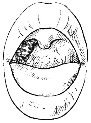
Pharyngeal membrane of diphtheria
Note: the membrane extends beyond the tonsils and covers the adjacent pharyngeal wall.
Treatment
Antitoxin
- ►
Give 40 000 U diphtheria antitoxin (IM or IV) immediately, because delay can increase the risk for mortality. As there is a small risk for a serious allergic reactionto the horse serum in the antitoxin, an initial intradermal test to detect hypersensitivity should be carried out, as described in the instructions, and treatment for anaphylaxis should be available.
Antibiotics
- ►
Any child with suspected diphtheria should be given a daily deep IM injection of procaine benzylpenicillin at 50 mg/kg (maximum, 1.2 g) daily for 10 days. This drug should not be given IV.
Oxygen
- ►
Avoid using oxygen unless there is incipient airway obstruction.
Signs such as severe lower chest wall indrawing and restlessness are more likely to indicate the need for tracheostomy (or intubation) than oxygen. Moreover, the use of a nasal or nasopharyngeal catheter can upset the child and precipitate obstruction of the airway.
- ►
However, oxygen should be given if there is incipient airway obstruction and intubation or a tracheostomy is deemed necessary.
Tracheostomy/intubation
- ►
Tracheostomy should be performed, only by experienced staff, if there are signs of incipient complete airway obstruction, such as severe lower chest wall indrawing and restlessness. If obstruction occurs, an emergency tracheostomy should be carried out. Orotracheal intubation is an alternative but may dislodge the membrane and fail to relieve the obstruction.
Supportive care
- ►
If the child has fever (≥ 39 or ≥ 102.2 °F) that appears to be causing distress, give paracetamol.
- ►
Encourage the child to eat and drink. If the child has difficulty in swallowing, nasogastric feeding is required. The nasogastric tube should be placed by an experienced clinician or, if available, an anaesthetist.

‘Bull neck’: a sign of diphtheria, due to enlarged lymph nodes in the neck
Avoid frequent examinations and invasive procedures when possible or disturbing the child unnecessarily.
Monitoring
The child's condition, especially respiratory status, should be assessed by a nurse every 3 h and by a doctor twice a day. The child should occupy a bed close to the nursing station, so that any sign of incipient airway obstruction can be detected as soon as it develops.
Complications
Myocarditis and paralysis may occur 2–7 weeks after the onset of illness.
- Signs of myocarditis include a weak, irregular pulse and evidence of heart failure. Refer to standard paediatric textbooks for details of the diagnosis and management of myocarditis.
Public health measures
- ►
The child should be nursed in a separate room by staff who are fully vaccinated against diphtheria.
- ►
Give all vaccinated household contacts a diphtheria toxoid booster.
- ►
Give all unvaccinated household contacts one dose of benzathine penicillin (600 000 U for those aged ≤ 5 years, 1 200 000 U for those > 5 years). Give them diphtheria toxoid, and check daily for 5 days for any signs of diphtheria.
4.6.3. Epiglottitis
Epiglottitis is a medical emergency that may result in death if not treated quickly. It is mainly caused by the bacteria H. influenzae type b but may also be caused by other bacteria or viruses associated with upper respiratory infections. Epiglottitis usually begins as an inflammation and swelling between the base of the tongue and the epiglottis. The swelling may obstruct the airway.
Diagnosis
- sore throat with difficulty in speaking
- difficulty in breathing
- soft stridor
- fever
- drooling of saliva
- difficulty in swallowing or inability to drink.
Treatment
Treatment of patients with epiglottitis is directed to relieving the airway obstruction and eradicating the infectious agent.
- ►
Keep the child calm, and provide humidified oxygen, with close monitoring.
- ►
Avoid examining the throat if the signs are typical, to avoid precipitating obstruction.
- ►
Call for help and secure the airway as an emergency because of the danger of sudden, unpredictable airway obstruction. Elective intubation is the best treatment if there is severe obstruction but may be very difficult; consider the need for surgical intervention to ensure airway patency.
- ►
Give IV antibiotics when the airway is safe: ceftriaxone at 80 mg/kg once daily for 5 days.
4.6.4. Anaphylaxis
Anaphylaxis is a severe allergic reaction, which may cause upper airway obstruction with stridor, lower airway obstruction with wheezing or shock or all three. Common causes include allergic reactions to antibiotics, to vaccines, to blood transfusion and to certain foods, especially nuts.
Consider the diagnosis if any of the following symptoms is present and there is a history of previous severe reaction, rapid progression or a history of asthma, eczema or atopy.
| Severity | Symptoms | signs |
|---|---|---|
| Mild |
|
|
| Moderate |
|
|
| Severe |
|
|
This situation is potentially life-threatening and may result in a change in level of consciousness, collapse, or respiratory or cardiac arrest.
- ►
Assess the airways, breathing and circulation.
- –
If the child is not breathing, give five rescue breaths with a bag-valve mask and 100% oxygen and assess circulation.
- –
If no pulse, start basic life support.
Treatment
- ►
Remove the allergen as appropriate.
- ►
For mild cases (just rash and itching), give oral antihistamine and oral prednisolone at 1 mg/kg.
- ►
For moderate cases with stridor and obstruction or wheeze:
- –
Give adrenaline at 0.15 ml of 1:1000 IM into the thigh (or subcutaneous); the dose may be repeated every 5–15 min.
- ►
For severe anaphylactic shock:
- –
Give adrenaline at 0.15 ml of 1:1000 IM and repeat every 5–15 min.
- –
Give 100% oxygen.
- –
Ensure stabilization of the airway, breathing, circulation and secure IV access.
- –
If the obstruction is severe, consider intubation or call an anaesthetist and surgeon to intubate or create a surgical airway.
- –
Administer 20 ml/kg normal saline 0.9% or Ringer's lactate solution IV as rapidly as possible. If IV access is not possible, insert an intraosseous line.
4.7. Conditions presenting with chronic cough
A chronic cough is one that lasts ≥ 14 days. Many conditions may present with a chronic cough such as TB, pertussis, foreign body or asthma (see Table 10).
Table 10Differential diagnosis in a child presenting with chronic cough
| Diagnosis | In favour |
|---|---|
| TB |
|
| Asthma |
|
| Foreign body |
|
| Pertussis |
|
| HIV |
|
| Bronchiectasis |
|
| Lung abscess |
|
History
- duration of coughing
- nocturnal cough
- paroxysmal cough or associated severe bouts ending with vomiting or whooping
- weight loss or failure to thrive (check growth chart, if available),
- night sweats
- persistent fever
- close contact with a known case of sputum-positive TB or pertussis
- history of attacks of wheeze and a family history of allergy or asthma
- history of choking or inhalation of a foreign body
- child suspected or known to be HIV-infected
- treatment given and response.
Examination
- fever
- lymphadenopathy (generalized and localized, e.g. in the neck)
- wasting
- wheeze or prolonged expiration
- clubbing
- apnoeic episodes (with pertussis)
- subconjunctival haemorrhages
- signs associated with foreign body aspiration:
- –
unilateral wheeze
- –
area of decreased breath sounds that is either dull or hyper-resonant on percussion
- –
deviation of the trachea or apex beat
- signs associated with HIV infection.
Treatment guidelines for the most common causes of chronic cough are indicated below:
- Asthma.
- Pertussis.
- TB.
- Foreign body.
- HIV.
4.7.1. Pertussis
Pertussis is most severe in young infants who have not yet been immunized. After an incubation period of 7–10 days, the child has fever, usually with a cough and nasal discharge that are clinically indistinguishable from the common cough and cold. In the second week, there is paroxysmal coughing that can be recognized as pertussis. The episodes of coughing can continue for 3 months or longer. The child is infectious for up to 3 weeks after the onset of bouts of whooping cough.
Diagnosis
Suspect pertussis if a child has had a severe cough for more than 2 weeks, especially if the disease is known to be occurring locally. The most useful diagnostic signs are:
- paroxysmal coughing followed by a whoop when breathing in, often with vomiting
- subconjunctival haemorrhages
- child not vaccinated against pertussis
- young infants may not whoop; instead, the cough may be followed by suspension of breathing (apnoea) or cyanosis, or apnoea may occur without coughing.
Also examine the child for signs of pneumonia, and ask about convulsions.
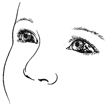
Subconjunctival haemorrhages prominent on the white sclera
Treatment
Treat mild cases in children aged ≥ 6 months at home with supportive care (see below). Admit infants aged < 6 months to hospital; also admit any child with pneumonia, convulsions, dehydration, severe malnutrition or prolonged apnoea or cyanosis after coughing.
Antibiotics
- ►
Give oral erythromycin (12.5 mg/kg four times a day) for 10 days. This does not shorten the illness but reduces the period of infectiousness.
- ►
Alternatively, if available, give azithromycin at 10 mg/kg (maximum, 500 mg) on the first day, then 5 mg/kg (maximum, 250 mg) once a day for 4 days.
- ►
If there is fever, if erythromycin or azithromycin is not available, or if there are signs of pneumonia, treat with amoxicillin as possible secondary pneumonia. Follow the other guidelines for severe pneumonia (see 4.2.1).
Oxygen
- ►
Give oxygen to children who have spells of apnoea or cyanosis, severe paroxysms of coughing or low oxygen saturation ≤ 90% on a pulse oximeter.
Use nasal prongs, not a nasopharyngeal catheter or nasal catheter, which can provoke coughing. Place the prongs just inside the nostrils and secure with a piece of tape just above the upper lip. Care should be taken to keep the nostrils clear of mucus, as this blocks the flow of oxygen. Set a flow rate of 1–2 litres/min (0.5 litre/min for young infants). Humidification is not required with nasal prongs.
- ►
Continue oxygen therapy until the above signs are no longer present, after which there is no value in continuing oxygen.
- ►
A nurse should check, every 3 h, that the prongs or catheter are in the correct place and not blocked with mucus and that all connections are secure. See section 10.7 for further details.
Airway management
- ►
During paroxysms of coughing, place the child in the recovery position to prevent inhalation of vomitus and to aid expectoration of secretions.
- –
If the child has cyanotic episodes, clear secretions from the nose and throat with brief, gentle suction.
- –
If apnoea occurs, clear the airways immediately with gentle suction under direct vision, breathe for the infant using a bag-valve mask ideally with a reservoir bag and connected to high-flow oxygen
Supportive care
- Avoid, as far as possible, any procedure that could trigger coughing, such as application of suction, throat examination or use of a nasogastric tube (unless the child cannot drink).
- Do not give cough suppressants, sedatives, mucolytic agents or antihistamines.
- ►
If the child has fever (≥ 39 °C, ≥ 102.2 °F) that appears to be causing distress, give paracetamol.
- ►
Encourage breastfeeding or oral fluids. If the child cannot drink, pass a nasogastric tube and give small, frequent amounts of fluid (ideally expressed breast milk) to meet the child's maintenance needs. If there is severe respiratory distress and maintenance fluids cannot be given through a nasogastric tube because of persistent vomiting, give IV fluids to avoid the risk of aspiration and avoid triggering coughing.
Ensure adequate nutrition by giving small, frequent feeds. If there is continued weight loss despite these measures, feed the child by nasogastric tube.
Monitoring
The child should be assessed by a nurse every 3 h and by a doctor once a day. To facilitate early detection and treatment of apnoeic or cyanotic spells or severe episodes of coughing, the child should occupy a bed in a place close to the nursing station, where oxygen and assisted ventilation are available. Also, teach the child's mother to recognize apnoeic spells and to alert the nurse if these occur.
Complications
Pneumonia: This is the commonest complication of pertussis and is caused by secondary bacterial infection or inhalation of vomit.
- ■
Signs suggesting pneumonia include fast breathing between coughing episodes, fever and the rapid onset of respiratory distress.
- ►
Treat pneumonia in children with pertussis as follows:
- –
Give parenteral ampicillin (or benzylpenicillin) and gentamicin for 5 days, or alternatively give azithromycin for 5 days.
- –
Give oxygen as described for the treatment of severe pneumonia (see sections 4.2.1 and 10.7).
Convulsions. These may result from anoxia associated with an apnoeic or cyanotic episode or toxin-mediated encephalopathy.
- ►
If a convulsion does not stop within 2 min, give diazepam, following the guidelines in Chapter 1 (Chart 9).
Malnutrition. Children with pertussis may become malnourished as a result of reduced food intake and frequent vomiting.
- ►
Prevent malnutrition by ensuring adequate feeding, as described above, under ‘Supportive care’.
Haemorrhage and hernias
- ■
Subconjunctival haemorrhage and epistaxis are common during pertussis.
- ►
No specific treatment is needed.
- ■
Umbilical or inguinal hernias may be caused by violent coughing.
- ►
Do not treat them unless there are signs of bowel obstruction, but refer the child for surgical evaluation after the acute phase.
Public health measures
- ►
Give DPT vaccine to any child in the family who is not fully immunized and to the child with pertussis.
- ►
Give a DPT booster to previously vaccinated children.
- ►
Give erythromycin estolate (12.5 mg/kg four times a day) for 10 days to any infant in the family who is < 6 months old and has fever or other signs of a respiratory infection.
4.7.2. Tuberculosis
Most children infected with M. tuberculosis do not develop TB. The only evidence of infection may be a positive skin test. The development of TB depends on the competence of the immune system to resist multiplication of the M. tuberculosis infection. This competence varies with age, being least in the very young. HIV infection and malnutrition lower the body's defenses, and measles and whooping cough temporarily impair the strength of the immune system. In the presence of any of these conditions, TB can develop more easily.
TB is most often severe when it is located in the lungs, meninges or kidney. Cervical lymph nodes, bones, joints, abdomen, ears, eyes and skin may also be affected. Many children present only with failure to grow normally, weight loss or prolonged fever. Cough for > 14 days can also be a presenting sign; in children, however, sputum-positive pulmonary TB is rarely diagnosed.
Diagnosis
The risk for TB is increased when there is an active case (infectious, smear-positive pulmonary TB) in the same house or when the child is malnourished, has HIV/AIDS or had measles in the past few months. Consider TB in any child with:
A history of:
- unexplained weight loss or failure to grow normally
- unexplained fever, especially when it continues for longer than 2 weeks
- chronic cough (i.e. cough for > 14 days, with or without a wheeze)
- exposure to an adult with probable or definite infectious pulmonary TB.
On examination:
- fluid on one side of the chest (reduced air entry, stony dullness to percussion)
- enlarged non-tender lymph nodes or a lymph node abscess, especially in the neck
- signs of meningitis, especially when these develop over several days and the spinal fluid contains mostly lymphocytes and elevated protein
- abdominal swelling, with or without palpable lumps
- progressive swelling or deformity in the bone or a joint, including the spine
Investigations
- Try to obtain specimens for microscopic examination of acid-fast bacilli (Ziehl-Neelsen stain) and for culture of tubercle bacilli. Possible specimens include three consecutive early-morning, fasting gastric aspirates, CSF (if clinically indicated) and pleural fluid and ascites fluid (if present). As the detection rates with these methods are low, a positive result confirms TB, but a negative result does not exclude the disease.
- New rapid diagnostic tests are more accurate and may be more widely available in future.
- Obtain a chest X-ray. A diagnosis of TB is supported when a chest X-ray shows a miliary pattern of infiltrates or a persistent area of infiltrate or consolidation, often with pleural effusion, or a primary complex.
- Perform a purified protein derivative skin test (PPD or mantoux test). The test is usually positive in children with pulmonary TB (reactions of > 10 mm suggest TB; < 10 mm in a child previously vaccinated with BCG is equivocal). The purified protein derivative test may be negative in children with TB who have HIV/AIDS, miliary disease, severe malnutrition or recent measles.
- Xpert MTB/RIF should be used as the initial diagnostic test in children suspected of having multidrug-resistant TB (MDR-TB) or HIV-associated TB.
- Routine HIV testing should be offered to all children suspected of TB.
Treatment
- ►
Give a full course of treatment to all confirmed or strongly suspected cases.
- ►
When in doubt, e.g. in a child with strongly suspected TB or who fails to respond to treatment for other probable diagnoses, give treatment for TB.
Treatment failures for other diagnoses include antibiotic treatment for apparent bacterial pneumonia (when the child has pulmonary symptoms), for possible meningitis (when the child has neurological symptoms) or for intestinal worms or giardiasis (when the child fails to thrive or has diarrhoea or abdominal symptoms).
- ►
Suspected or confirmed childhood TB should be treated with a combination of anti-TB drugs, depending on the severity of disease, HIV status and level of isoniazid resistance.
- ►
Follow the national TB programme guidelines for recommended treatment.
- ►
To reduce the risk for drug-induced hepatotoxicity in children, follow the recommended dosages:
- –
Isoniazid (H): 10 mg/kg (range, 10–15 mg/kg); maximum dose, 300 mg/day
- –
Rifampicin (R): 15 mg/kg (range, 10–20 mg/kg); maximum dose, 600 mg/kg per day
- –
Pyrazinamide (Z): 35 mg/kg (range, 30–40 mg/kg)
- –
Ethambutol (E): 20 mg/kg (range, 15–25 mg/kg).
Treatment regimens
If national recommendations are not available, follow the WHO guidelines according to the regimens given below:
- ►
Four-drug regimen: HRZE for 2 months, followed by a two-drug (HR) regimen for 4 months for all children with suspected or confirmed pulmonary TB or peripheral lymphadenitis living in an area of high HIV prevalence or where resistance to H is high or children with extensive pulmonary disease living in areas of low HIV prevalence or low H resistance;
- ►
Three-drug regimen: HRZ for 2 months, followed by a two-drug (HR) regimen for 4 months for children with suspected or confirmed pulmonary TB or tuberculous peripheral lymphadenitis living in areas of low HIV prevalence or low H resistance or HIV-negative;
- ►
In cases of suspected or confirmed tuberculous meningitis, spinal TB with neurological signs or osteo-articular TB, treat for 12 months with a four-drug regimen (HRZE) for 2 months, followed by a two-drug (HR) regimen for 10 months;
- ►
In infants (aged 0–3 months) with suspected or confirmed pulmonary TB or tuberculous peripheral lymphadenitis, treat promptly with the standard regimens described above, with adjustment of doses to reconcile the effect of age and possible toxicity in young infants.
Intermittent regimens: In areas with well-established directly observed therapy, thrice-weekly regimens can be considered for children known to be HIV-negative. They should not be used in areas with a high HIV prevalence, because there is a high risk of treatment failure and development of multidrug-resistant TB.
Precautions: Streptomycin should not be used as part of first-line treatment regimens for children with pulmonary TB or tuberculous peripheral lymphadenitis. It should be reserved for the treatment of multidrug-resistant TB in children with known susceptibility to this medicine.
Multidrug-resistant TB
- ►
In cases of MDR TB, treat children with proven or suspected pulmonary TB or tuberculous meningitis with a fluoroquinolone or other second-line TB drug. An appropriate MDR TB treatment regimen in the context of a well-functioning MDR TB control programme should be used. The decision to treat should be taken by a clinician experienced in managing paediatric TB.
Monitoring
Confirm that the medication is being taken as instructed, by direct observation of each dose. Monitor the child's weight gain daily and temperature twice a day in order to check for resolution of fever. These are signs of response to therapy. When treatment is given for suspected TB, improvement should be seen within 1 month. If this does not occur, review the patient, check compliance, re-investigate and reconsider the diagnosis.
Public health measures
- ►
Notify the case to the responsible district health authorities. Ensure that treatment is monitored as recommended by the national TB programme. Check all household members of the child (and, if necessary, school contacts) for undetected cases of TB, and arrange treatment for any that are found.
- ►
Children < 5 years of age who are household or close contacts of people with TB and who, after an appropriate clinical evaluation, are found not to have active TB should be given 6 months of isoniazid preventive therapy (10 mg/kg/day, range 7–15 mg/kg, maximum dose 300 mg/day).
Follow-up
A programme of ‘active’ follow-up, in which a health worker visits the child and his or her family at home, can reduce default from TB treatment. During follow-up at home or in hospital, health workers can:
- Check whether medications for TB are being taken regularly.
- Remind the family and the treatment supporter about the importance of taking medications regularly, even if the child is well, for the full duration of treatment.
- Screen family contacts, including other children in the family, by inquiring about cough, and start these children on isoniazid preventive therapy.
- Suggest how the family's home environment might be made healthier for children, such as eliminating smoking inside the house, good ventilation and hand-washing.
- Discuss with the parents the importance of nutrition in recovery from TB and any problems in providing good nutrition for their children.
- Check the child for growth, nutritional state and signs of TB and other treatable conditions. If problems are found, the health worker should recommend how these can be treated or refer the family to a paediatrician.
- Check the child's health record, and tell the parents when and where they should bring the child for doses of vaccine.
- Ask the parents if they have any questions or concerns, and answer or discuss these, or refer the family to a paediatrician.
- Record their observations on the TB treatment card.
4.7.3. Foreign body inhalation
Nuts, seeds or other small objects may be inhaled, most often by children < 4 years of age. The foreign body usually lodges in a bronchus (more often in the right) and can cause collapse or consolidation of the portion of lung distal to the site of blockage. Choking is a frequent initial symptom. This may be followed by a symptom-free interval of days or weeks before the child presents with persistent wheeze, chronic cough or pneumonia, which fails to respond to treatment. Small sharp objects can lodge in the larynx, causing stridor or wheeze. Rarely, a large object lodged in the larynx can cause sudden death from asphyxia, unless it can be dislodged or an emergency tracheostomy be done.
Diagnosis
Inhalation of a foreign body should be considered in a child with the following signs:
- sudden onset of choking, coughing or wheezing; or
- segmental or lobar pneumonia that fails to respond to antibiotic therapy.
Examine the child for:
- unilateral wheeze
- an area of decreased breath sounds that is either dull or hyper-resonant on percussion
- deviation of the trachea or apex beat.
Obtain a chest X-ray at full expiration to detect an area of hyperinflation or collapse, mediastinal shift (away from the affected side) or a foreign body if it is radio-opaque.
Treatment
Emergency first aid for the choking child: Attempt to dislodge and expel the foreign body. The management depends on the age of the child.
For infants
- ►
Lay the infant in a head-down position on one of your arms or on your thigh.
- ►
Strike the middle of the infant's back five times with the heel of your hand.
- ►
If the obstruction persists, turn the infant over and give five firm chest thrusts with two fingers on the lower half of the sternum.
- ►
If the obstruction persists, check the infant's mouth for any obstruction that can be removed.
- ►
If necessary, repeat this sequence with back slaps.
For older children
- ►
While the child is sitting, kneeling or lying, strike the child's back five times with the heel of the hand.
- ►
If the obstruction persists, go behind the child and pass your arms around the child's body; form a fist with one hand immediately below the sternum; place the other hand over the fist, and thrust sharply upwards into the abdomen. Repeat this up to five times.
- ►
Then check the child's mouth for any obstruction that can be removed.
- ►
If necessary, repeat the sequence with back slaps.
Once this has been done, it is important to check the patency of the airway by:
- looking for chest movements
- listening for breath sounds and
- feeling for breath.
If further management of the airways is required after the obstruction is removed, Chart 4, describes actions that will keep the child's airways open and prevent the tongue from falling back to obstruct the pharynx while the child recovers.
- ►
Later treatment of suspected foreign body aspiration. If a foreign body is suspected, refer the child to a hospital where diagnosis is possible and the object can be removed after bronchoscopy. If there is evidence of pneumonia, begin treatment with ampicillin (or benzylpenicillin) and gentamicin, as for severe pneumonia, before attempting to remove the foreign body.
4.8. Heart failure
Heart failure causes fast breathing and respiratory distress. The underlying causes include congenital heart disease (usually in the first months of life), acute rheumatic fever, cardiac arrhythmia, myocarditis, suppurative pericarditis with constriction, infective endocarditis, acute glomerulonephritis, severe anaemia, severe pneumonia and severe malnutrition. Heart failure can be precipitated or worsened by fluid overload, especially when large volumes of IV fluids are given.
Diagnosis
The commonest signs of heart failure, on examination, are:
- tachycardia (heart rate > 160/min in a child < 12 months; > 120/min in a child aged 12 months to 5 years)
- gallop rhythm with basal crackles on auscultation
- enlarged, tender liver
- in infants, fast breathing (or sweating), especially when feeding (see section 4.1, for definition of fast breathing); in older children, oedema of the feet, hands or face or distended neck veins (raised jugular venous pressure)

Raised jugular venous pressure – a sign of heart failure
Severe palmar pallor may be present if severe anaemia is the cause of the heart failure.
Heart murmur may be present in rheumatic heart disease, congenital heart disease or endocarditis.
If the diagnosis is in doubt, a chest X-ray can be taken and may show an enlarged heart or abnormal shape.
Measure blood pressure if possible. If it is raised, consider acute glomerulonephritis (See standard paediatric textbook for treatment).
Treatment
Treatment depends on the underlying heart disease (Consult international or national paediatric guidelines). The main measures for treating heart failure in children who are not severely malnourished are:
- ►
Oxygen. Give oxygen if the child has a respiratory rate of ≥ 70/min, shows signs of respiratory distress, or has central cyanosis or low oxygen saturation. Aim to keep oxygen saturation > 90%. See section 10.7.
- ►
Diuretics. Give furosemide: A dose of 1 mg/kg should increase urine flow within 2 h. For faster action, give the drug IV. If the initial dose is not effective, give 2 mg/kg and repeat in 12 h, if necessary. Thereafter, a single daily dose of 1–2 mg/kg orally is usually sufficient.
- ►
Digoxin. Consider giving digoxin (see Annex 2).
- ►
Supplemental potassium. Supplemental potassium is not required when furosemide is given alone for treatment lasting only a few days. When digoxin and furosemide are given, or if furosemide is given for more than 5 days, give oral potassium at 3–5 mmol/kg per day.
Supportive care
- Avoid giving IV fluids, if possible.
- Support the child in a semi-seated position with head and shoulders elevated and lower limbs dependent.
- Relieve any fever with paracetamol to reduce the cardiac workload.
- Consider transfusion if severe anaemia is present.
Monitoring
The child should be checked by a nurse every 6 h (every 3 h while on oxygen therapy) and by a doctor once a day. Monitor both respiratory and pulse rates, liver size and body weight to assess the response to treatment. Continue treatment until the respiratory and pulse rates are normal and the liver is no longer enlarged.
4.9. Rheumatic heart disease
Chronic rheumatic heart disease is a complication of acute rheumatic fever, which leaves permanent damage to the heart valves. In some children, antibodies produced in response to group A β-haemolytic streptococci lead to varying degrees of pancarditis, with associated valve insufficiency in the acute phase.
The risk for rheumatic heart disease is higher with repeated episodes of acute rheumatic fever. It leads to valve stenosis, with varying degrees of regurgitation, atrial dilatation, arrhythmia and ventricular dysfunction. Chronic rheumatic heart disease is a major cause of mitral valve stenosis in children.
Diagnosis
Rheumatic heart disease should be suspected in any child with a previous history of rheumatic fever who presents with heart failure or is found to have a heart murmur. Diagnosis is important because penicillin prophylaxis can prevent further episodes of rheumatic fever and avoid worse damage to the heart valves.
The presentation depends on the severity. Mild disease may cause few symptoms except for a heart murmur in an otherwise well child and is rarely diagnosed. Severe disease may present with symptoms that depend on the extent of heart damage or the presence of infective endocarditis.
History
- chest pain
- heart palpitations
- symptoms of heart failure (including orthopnoea, paroxysmal nocturnal dyspnoea and oedema)
- fever or stroke usually associated with infection of damaged heart valves
- breathlessness on exertion or exercise
- fainting (syncope)
Examination
- signs of heart failure
- cardiomegaly with a heart murmur
- signs of infective endocarditis (e.g. conjunctival or retinal haemorrhages, hemiparesis, Osler nodes, Roth spots and splenomegaly)
Investigations
- chest X-ray: cardiomegaly with congested lungs
- an echocardiogram, if available, is useful for confirming rheumatic heart disease, the extent of valve damage and evidence of infective endocarditis.
- full blood count
- blood culture
Management
- Admit the child if in heart failure or has suspected bacterial endocarditis.
- Treatment depends on the type and extent of valvular damage.
- Manage heart failure if present.
- ►
Give diuretics to relieve symptoms of pulmonary congestion and vasodilators when necessary.
- ►
Give penicillin or ampicillin or ceftriaxone plus gentamicin IV or IM for 4–6 weeks for infective endocarditis.
- ►
Refer for echocardiographic evaluation and decision on long-term management. May require surgical management in severe valvular stenosis or regurgitation.
Follow-up care
- All children with rheumatic heart disease should receive routine antibiotic prophylaxis.
- ►
Give benzathine benzylpenicillin at 600 000 U IM every 3–4 weeks
- Ensure antibiotic prophylaxis for endocarditis before dental and invasive surgical procedures.
- Ensure that vaccinations are up to date.
- Review every 3–6 months, depending on severity of valvular damage.
Complications
Infective endocarditis is more common. It presents with fever and heart murmur in a very unwell child. Treat with ampicillin and gentamicin for 6 weeks.
Atrial fibrillation or thromboembolism may occur, especially in the presence of mitral stenosis.
- Cough or difficulty in breathing - Pocket Book of Hospital Care for ChildrenCough or difficulty in breathing - Pocket Book of Hospital Care for Children
- FADS1 [Apteryx rowi]FADS1 [Apteryx rowi]Gene ID:112978023Gene
- RecName: Full=Muscarinic acetylcholine receptor M1RecName: Full=Muscarinic acetylcholine receptor M1gi|113118|sp|P11229.2|ACM1_HUMANProtein
- GMPPB [Mauremys reevesii]GMPPB [Mauremys reevesii]Gene ID:120409294Gene
Your browsing activity is empty.
Activity recording is turned off.
See more...