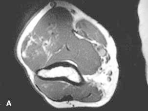
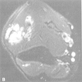
Figure 36e-4. T1-weighted (A) and T2-weighted, fat suppressed (B) axial MR images through the upper arm reveal a lobulated mass with interdigitating fat signal and extension through fascial planes.

Figure 36e-4

T1-weighted (A) and T2-weighted, fat suppressed (B) axial MR images through the upper arm reveal a lobulated mass with interdigitating fat signal and extension through fascial planes. These characteristics are diagnostic of a soft tissue hemangioma.

From: Chapter 36e, Imaging of Musculoskeletal Neoplasms
Cover of Holland-Frei Cancer Medicine
Holland-Frei Cancer Medicine. 6th edition.
Kufe DW, Pollock RE, Weichselbaum RR, et al., editors.
Hamilton (ON): BC Decker; 2003.
Copyright © 2003, BC Decker Inc.

NCBI Bookshelf. A service of the National Library of Medicine, National Institutes of Health.