From: Chapter 36e, Imaging of Musculoskeletal Neoplasms
NCBI Bookshelf. A service of the National Library of Medicine, National Institutes of Health.
Kufe DW, Pollock RE, Weichselbaum RR, et al., editors. Holland-Frei Cancer Medicine. 6th edition. Hamilton (ON): BC Decker; 2003.

Holland-Frei Cancer Medicine. 6th edition.
Show details

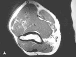
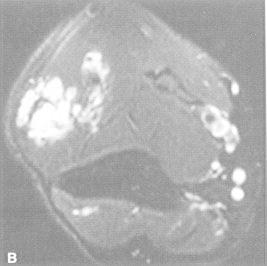
Figure 36e-4
T1-weighted (A) and T2-weighted, fat suppressed (B) axial MR images through the upper arm reveal a lobulated mass with interdigitating fat signal and extension through fascial planes. These characteristics are diagnostic of a soft tissue hemangioma.
- Figure 36e-4, [T1-weighted (A) and T2-weighted, fat...]. - Holland-Frei Cancer M...Figure 36e-4, [T1-weighted (A) and T2-weighted, fat...]. - Holland-Frei Cancer Medicine
- Danio rerio ovarian cytochrome P450 aromatase (cyp19a) mRNA, complete cdsDanio rerio ovarian cytochrome P450 aromatase (cyp19a) mRNA, complete cdsgi|12655891|gb|AF226620.1|Nucleotide
- cyclin-dependent kinase-like 3 isoform 3 [Homo sapiens]cyclin-dependent kinase-like 3 isoform 3 [Homo sapiens]gi|664806087|ref|NP_001287782.1|Protein
- trip12 thyroid hormone receptor interactor 12 [Danio rerio]trip12 thyroid hormone receptor interactor 12 [Danio rerio]Gene ID:564866Gene
Your browsing activity is empty.
Activity recording is turned off.
See more...