Summary
The purpose of this overview on hereditary ataxia is to increase the awareness of clinicians regarding the causes of hereditary ataxia, related genetic counseling issues, and management.
Goal 1.
Briefly describe the clinical characteristics of hereditary ataxias (sometimes referred to as "primary hereditary ataxias") for which an adult with ataxia or the caregivers of a child with ataxia would seek diagnosis and management from a neurologist as part of a multidisciplinary team.
Goal 2.
Review common and notable genetic causes of hereditary ataxia.
Goal 3.
Provide an evaluation strategy to identify the genetic cause of hereditary ataxia in a proband.
Goal 4.
Inform genetic counseling of family members of an individual with hereditary ataxia.
Goal 5.
Review management of hereditary ataxia.
1. Clinical Characteristics of Primary Hereditary Ataxia
For the purposes of this chapter, which deals exclusively with hereditary ataxias, the term "primary hereditary ataxia" has been used to designate hereditary ataxias for which an adult with ataxia or the caregivers of a child with ataxia would seek diagnosis and management from a neurologist as part of a multidisciplinary team.
Use of the term "primary hereditary ataxia" is intended to exclude hereditary multisystem disorders in which ataxia may be observed, but is usually not the primary presenting manifestation.
Excluded Categories
For the purposes of this overview the following categories of hereditary disorders in which ataxia may occur are not considered primary hereditary ataxias:
- Infantile-onset multisystem disorder
- Epileptic encephalopathy
- Presence of distinctive MRI findings:
- Brain malformation (e.g., cerebellar hypoplasia, cerebellar vermis hypoplasia as in Joubert syndrome and related disorders)
- Leukodystrophy
- Developmental delay / intellectual disability
- An inborn error of metabolism (e.g., peroxisomal biogenesis disorders, Zellweger spectrum disorders, disorders of glycosylation)
- Primary mitochondrial disorders (See Primary Mitochondrial Disorders Overview.)
- Other complex multisystem disorders such as Niemann-Pick disease type C and late-onset Tay-Sachs disease (see HEXA Disorders), which can on occasion present with ataxia
Manifestations
The manifestations of many of the more common primary hereditary ataxias discussed in Section 2 of this overview become evident between ages 30 and 50 years, although manifestations of other ataxias are evident before age 25 years (e.g., ataxia with oculomotor apraxia, Friedreich ataxia) or before age five years (e.g., ataxia-telangiectasia).
The first manifestation of ataxia can include:
- Most commonly, a slowly progressive gait disorder that appears unsteady and predisposes to unexpected falls;
- Disequilibrium ("dizziness"), which may lead to an evaluation for peripheral vestibular dysfunction;
- Hand and finger clumsiness or tremor, which may raise the possibility of essential tremor or even parkinsonism;
- Slurring of speech or unexpected choking, which could lead to an evaluation for amyotrophic lateral sclerosis (ALS);
- Rarely, double vision, which could lead to an evaluation by an optometrist or ophthalmologist.
At disease onset, these manifestations may be intermittent or evident only at certain times (e.g., later in the day, when tired, after consuming alcohol). The manifestations typically become constant and slowly worsen.
Typical cerebellar features on neurologic examination include:
- Wide-based, staggering walk with difficulty performing tandem gait;
- Truncal instability when sitting unsupported;
- Difficulty with target maneuvers of the upper extremities (dysmetria, terminal tremor);
- Slowed rapid alternating movements (dysdiadochokinesis);
- Dysarthria (slowed or slurred articulation, variable pitch and loudness, monotonous or "scanning" speech);
- Abnormal eye movements (saccade intrusions in primary gaze, nystagmus in horizontal or vertical gaze, saccade hypermetria).
Non-cerebellar findings that can mimic or exacerbate the cerebellar ataxia (which can be identified by examination or testing) include:
- Alterations in descending frontal or parietal motor pathways (e.g., in normal pressure hydrocephalus);
- Brain stem changes that disrupt cerebellar pathways (e.g., in central vestibular dysfunction);
- Sensory pathway dysfunctions that alter input to the cerebellum (visual, peripheral vestibular, posterior column, peripheral sensory);
- Other sources of motor change, especially as they affect gait (weakness, rigidity, spasticity);
- Non-neurologic disorders (e.g., joint disease).
Brain imaging (MRI, MRS, PET) confirms the presence of cerebellar atrophy or hypoplasia.
Electronystagmography can document dysfunction in cerebellar, vestibular, or oculomotor pathways.
Progression of a hereditary ataxia usually leads to:
- Use of assistive devices for ambulation five to ten years after onset and ultimately to wheelchair dependence;
- Choking or falls resulting in, for example, head injury or hip fracture, which are common causes of morbidity and mortality;
- Infection and sepsis (from aspiration or other pneumonia, urinary tract infection, decubiti), especially prominent in the later stages of disease;
- Decline in self-care ability, increasing risk of falls, dependence on a feeding tube, and/or incontinence;
- The family or caregiver's need to consider more in-home care assistance or out-of-home placement.
Affected individuals do not usually live longer than 25 years after manifestations emerge.
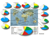
See Figure 1 for worldwide distribution of the most common primary hereditary ataxias.
2. Causes of Hereditary Ataxia
Note: Up to 40% of adults with late-onset cerebellar ataxia and no family history of ataxia will not have an identified genetic cause despite a comprehensive evaluation (see Section 3).
The causes of primary hereditary ataxia included in this overview are separated into nucleotide repeat disorders (Table 1 and Table 2), other common hereditary ataxias (Table 3), and potentially treatable causes of hereditary ataxia (Table 4 and Table 5).
Nucleotide Repeat Disorders
The nucleotide repeat disorders (see Table 1), the most common cause of hereditary ataxia, are discussed separately because of their unique molecular mechanism and inheritance issues.
Molecular Mechanism
In nucleotide repeat disorders, a sequence of nucleotides is repeated a number of times in tandem within a gene (in an exon or intron) or near a gene. For a given gene, the size of the nucleotide repeats varies: smaller numbers of repeats are common and not associated with phenotypic abnormalities, whereas abnormally large numbers of repeats (uninterrupted or interrupted) may be associated with phenotypic abnormalities.
Inheritance Issues
All three modes of inheritance can be observed in nucleotide repeat disorders: autosomal dominant, autosomal recessive, and X-linked.
Autosomal dominant inheritance. A unique aspect of autosomal dominant inheritance of nucleotide repeat disorders is anticipation, the earlier onset and increasing severity of disease in subsequent generations as a result of expansion in the repeat size during transmission. In some disorders, anticipation may be so extreme that children with early-onset, severe, and usually phenotypically different disease die of disease complications long before the affected parent or grandparent is symptomatic.
X-linked inheritance. A unique aspect of fragile X tremor/ataxia syndrome, the most common X-linked ataxia, is its occurrence in males and females with repeat sizes in the premutation range (see Table 2).
Table 1.
Hereditary Ataxias Caused by Nucleotide Repeat Expansions: Clinical Findings
Table 2.
Hereditary Ataxias Caused by Nucleotide Repeat Expansions: Molecular Genetics
Other Common Hereditary Ataxias
Table 3.
Most Common Hereditary Ataxias (Excluding Nucleotide Repeat Disorders)
3. Evaluation Strategies to Identify the Genetic Cause of Hereditary Ataxia in a Proband
Establishing a specific genetic cause of primary hereditary ataxia (as defined in this chapter):
- Can aid in discussions of prognosis (which are beyond the scope of this GeneReview) and genetic counseling;
- Usually involves a medical history, physical examination, family history, and genomic/genetic testing.
Medical History
Most of the common primary hereditary ataxias start similarly with an unsteady gait, imbalance or "dizziness," unexpected falls, clumsiness, and tremors.
Distinctive features of the medical history that could suggest a specific diagnosis (see Table 1 and Table 3) include the following:
- Age of onset:
- After age 50 years: SCA6
- Before age 20 years: Friedreich ataxia, ataxia with oculomotor apraxia types 1 and 2
- Before age five years: ataxia-telangiectasia
- Infantile: SCA2, SCA7
- Onset with episodic features: SCA6, the episodic ataxias
- Associated with:
- Retinopathy: SCA7
- Seizure disorder: SCA10, infantile-onset SCA7, DRPLA
- Dementia: SCA2, SCA17, DRPLA
- Severe dizziness or vertigo: SCA3, SCA6, RFC1 CANVAS / spectrum disorder
- Muscle cramping: SCA2, SCA3
- Scoliosis, pes cavus, cardiomyopathy: Friedreich ataxia
- Immunodeficiency or cancer: ataxia-telangiectasia
Physical Examination
All the primary hereditary ataxias have cerebellar features, but some have specific cerebellar or extracerebellar changes on examination that can suggest a specific diagnosis (see Table 1 and Table 3). In addition to the information provided in the medical history above, the following may be observed:
- Early presence of slowed oculomotor saccades: SCA2, SCA7
- Ophthalmoplegia: SCA1, SCA2, SCA3
- Oculomotor apraxia: ataxia-telangiectasia, ataxia with oculomotor apraxia types 1 and 2
- Fixation instability (saccade intrusions) in primary gaze: Friedreich ataxia
- Ocular conjunctival and skin telangiectases: ataxia-telangiectasia
- Downbeat nystagmus: SCA6 and episodic ataxia type 2
- Central or peripheral vestibular involvement: SCA3, SCA6, RFC1 CANVAS / spectrum disorder
- Motor unit fasciculations: SCA2, SCA3
- Peripheral neuropathy: SCA3, RFC1 CANVAS / spectrum disorder
- Spasticity: SCA1, SCA3, SCA7
- Extrapyramidal signs: SCA1, SCA2, SCA3, SCA17, DRPLA
- Absent deep tendon reflexes and upgoing toes: Friedreich ataxia
Family History
A three-generation family history should be taken with attention to relatives with manifestations of hereditary ataxia and documentation of relevant findings through direct examination or review of medical records, including results of molecular genetic testing, neuroimaging studies, and autopsy examinations. Findings in the family that may assist in narrowing the scope of relevant hereditary ataxias include the following:
- Earlier onset and increasing severity of disease in subsequent generations suggest an autosomal dominant nucleotide repeat disorder associated with anticipation (see Table 1).
- An affected parent or grandparent suggests autosomal dominant inheritance.
- No male-to-male transmission of the disorder suggests X-linked inheritance.
- Affected sibs or consanguinity suggests autosomal recessive inheritance. Note: In communities with a high prevalence of an autosomal recessive ataxia (e.g., the ARSACS carrier frequency in the Saguenay–Lac-Saint-Jean region of Quebec is 1:21), affected individuals in two or more generations may be observed.
- Late-onset cerebellar ataxia in a grandfather who has a grandson with intellectual disability suggests fragile X-associated tremor/ataxia syndrome in the grandfather.
- Note: In the absence of a molecularly confirmed ataxia, reports of balance problems in a grandparent, parent, or sib do not necessarily indicate a shared genetic cause. Multifactorial and acquired cerebellar disorders, which are four to five times more common than inherited ataxias, can confuse a family history.
Molecular Genetic Testing
Nucleotide repeat disorders. Establishing the diagnosis in an individual with one of the nucleotide repeat disorders (see Table 1) requires identification of an expanded nucleotide repeat and determination of the nucleotide repeat size for each disorder (see Table 2).
Note that commercially available multigene panels that rely on sequence analysis alone will not identify these nucleotide repeat expansions; thus, specific assays are required to analyze the nucleotide repeat in each gene of interest. Options offered by some laboratories:
- A multigene "repeat expansion" panel to specifically identify nucleotide repeat expansions
- A multigene ataxia panel that combines both repeat expansion testing and sequence-based testing analysis
Non-nucleotide repeat disorders. Establishing the diagnosis of one of the disorders listed in Table 3 requires detection of a sequence variant. Molecular genetic testing approaches can include a combination of gene-targeted testing (multigene panel) and comprehensive genomic testing (exome sequencing, genome sequencing). Gene-targeted testing requires the clinician to hypothesize which gene(s) are likely involved, whereas genomic testing does not.
- A "sequence-based" ataxia multigene panel is most likely to identify the genetic cause of the condition while limiting identification of variants of uncertain significance and pathogenic variants in genes that do not explain the underlying phenotype. Note: (1) The genes included in the panel and the diagnostic sensitivity of the testing used for each gene vary by laboratory and are likely to change over time. (2) In some laboratories, panel options may include a custom laboratory-designed panel and/or custom phenotype-focused exome analysis that includes genes specified by the clinician. (3) Methods used in a panel may include sequence analysis, deletion/duplication analysis, and/or other non-sequencing-based tests. (4) Sequencing-based tests including exome sequencing do not readily detect nucleotide repeat expansions.
- Comprehensive genomic testing is an option that does not require the clinician to determine which gene(s) are likely involved. Exome sequencing is most commonly used; genome sequencing is also possible. Note that sequencing-based tests including exome sequencing do not readily detect nucleotide repeat expansions.
4. Genetic Counseling of Family Members of an Individual with Hereditary Ataxia
Genetic counseling is the process of providing individuals and families with information on the nature, mode(s) of inheritance, and implications of genetic disorders to help them make informed medical and personal decisions. The following section deals with genetic risk assessment and the use of family history and genetic testing to clarify genetic status for family members; it is not meant to address all personal, cultural, or ethical issues that may arise or to substitute for consultation with a genetics professional. —ED.
Mode of Inheritance
The hereditary ataxias included in this overview can be inherited in an autosomal dominant, autosomal recessive, or X-linked manner. Genetic counseling and risk assessment depend on determination of the specific cause of an inherited ataxia in an individual.
For hereditary ataxias that are nucleotide repeat disorders, select the relevant link to the GeneReview chapter (if available) in Table 1 for genetic counseling issues.
The genetic counseling issues for the other common hereditary ataxias (see Table 3) are discussed below.
Autosomal Dominant Inheritance – Risk to Family Members
Parents of a proband
- Most individuals diagnosed with autosomal dominant hereditary ataxia have an affected parent.
- Some individuals diagnosed with hereditary ataxia have the disorder as the result of a de novo pathogenic variant.
- Molecular genetic testing is recommended for the parents of a proband with an apparent de novo pathogenic variant.
- If the pathogenic variant identified in the proband is not identified in either parent and parental identity testing has confirmed biological maternity and paternity, the following possibilities should be considered:
- The proband has a de novo pathogenic variant.
- The proband inherited a pathogenic variant from a parent with germline (or somatic and germline) mosaicism. Note: Testing of parental leukocyte DNA may not detect all instances of somatic mosaicism and will not detect a pathogenic variant that is present in the germ cells only.
- The family history of some individuals diagnosed with autosomal dominant hereditary ataxia may appear to be negative because of failure to recognize the disorder in family members, early death of the parent before onset of symptoms, or late onset of the disease in the affected parent. Therefore, an apparently negative family history cannot be confirmed unless molecular genetic testing has demonstrated that neither parent is heterozygous for the pathogenic variant identified in the proband.
Sibs of a proband. The risk to the sibs of the proband depends on the clinical/genetic status of the proband's parents:
- If a parent of the proband is affected and/or is known to have the pathogenic variant identified in the proband, the risk to sibs is 50%.
- If the pathogenic variant found in the proband cannot be detected in the leukocyte DNA of either parent, the recurrence risk to sibs is slightly greater than that of the general population because of the possibility of parental germline mosaicism. (Note: Parental mosaicism has been reported in SCA29 [Ngo et al 2019] and other rarer ataxias.)
- If the parents have not been tested for the pathogenic variant but are clinically unaffected, sibs are still presumed to be at increased risk for hereditary ataxia because of the possibility of age-related penetrance in a heterozygous parent or the possibility of parental germline mosaicism.
Offspring of a proband. Each child of an individual with autosomal dominant hereditary ataxia has a 50% chance of inheriting the pathogenic variant.
Other family members. The risk to other family members depends on the status of the proband's parents: if a parent has the pathogenic variant, the parent's family members may be at risk.
Autosomal Recessive Inheritance – Risk to Family Members
Parents of a proband
- The parents of an individual diagnosed with autosomal recessive hereditary ataxia are presumed to be heterozygous for a pathogenic variant.
- Molecular genetic testing is recommended for the parents of a proband to confirm that both parents are heterozygous for a pathogenic variant and to allow reliable recurrence risk assessment.
- If a pathogenic variant is detected in only one parent and parental identity testing has confirmed biological maternity and paternity, it is possible that one of the pathogenic variants identified in the proband occurred as a de novo event in the proband or as a postzygotic de novo event in a mosaic parent [Jónsson et al 2017]. If the proband appears to have homozygous pathogenic variants (i.e., the same two pathogenic variants), additional possibilities to consider include:
- A single- or multiexon deletion in the proband that was not detected by sequence analysis and that resulted in the artifactual appearance of homozygosity;
- Uniparental isodisomy for the parental chromosome with the pathogenic variant that resulted in homozygosity for the pathogenic variant in the proband.
- Heterozygotes (carriers) are not at risk of developing autosomal recessive hereditary ataxia. (Note: Individuals who are heterozygous for an ATM pathogenic variant are at increased risk for cancer and coronary artery disease; see Ataxia-Telangiectasia.)
Sibs of a proband
- If both parents are known to be heterozygous for a pathogenic variant, each sib of an affected individual has at conception a 25% chance of being affected, a 50% chance of being heterozygous, and a 25% chance of inheriting neither of the familial pathogenic variants.
- Heterozygotes (carriers) are not at risk of developing autosomal recessive hereditary ataxia. (Note: Individuals who are heterozygous for an ATM pathogenic variant are at increased risk for cancer and coronary artery disease; see Ataxia-Telangiectasia.)
Offspring of a proband. The offspring of an individual with autosomal recessive hereditary ataxia are obligate heterozygotes (carriers) for a pathogenic variant.
Other family members. Each sib of the proband's parents is at a 50% risk of being a carrier of a pathogenic variant.
Carrier detection. Carrier testing for at-risk relatives requires prior identification of the ataxia-related pathogenic variants in the family.
Resources
GeneReviews staff has selected the following disease-specific and/or umbrella support organizations and/or registries for the benefit of individuals with this disorder and their families. GeneReviews is not responsible for the information provided by other organizations. For information on selection criteria, click here.
- Ataxia UKUnited KingdomPhone: 0800 995 6037; +44 (0) 20 7582 1444 (from abroad)Email: help@ataxia.org.uk
- National Ataxia FoundationPhone: 763-553-0020Fax: 763-553-0167Email: naf@ataxia.org
- Spanish Ataxia Federation (FEDAES)SpainPhone: 601 037 982Email: info@fedaes.org
- Associazione Italiana per la lotta alle Sindromi Atassiche (AISA)Via Sara 1216039ItalyPhone: 39 342 9124574Email: nazionale@atassia.it
- A-T Children's ProjectAtaxia-Telangiectasia Children's ProjectPhone: 800.5.HELP.A-T (800.543.5728); 954-481-6611
- euro-ATAXIA (European Federation of Hereditary Ataxias)United KingdomEmail: lporter@ataxia.org.uk
- FARAFriedreich's Ataxia Research AlliancePhone: 484-879-6160Fax: 484-872-1402Email: info@CureFA.org
- NCBI Genes and Disease
5. Management of Hereditary Ataxia
Evaluations Following Initial Diagnosis
To establish the extent of disease and needs of an individual diagnosed with a hereditary ataxia, the evaluations summarized in Table 6 (if not performed as part of the evaluation that led to the diagnosis) are recommended.
Table 6.
Recommended Evaluations Following Initial Diagnosis in Individuals with a Hereditary Ataxia
Treatment of Manifestations
The goals of supportive care are to maximize function and reduce complications. Depending on the clinical manifestations, affected individuals benefit from supportive care by a multidisciplinary team of specialists including neurologists, occupational therapists, physical therapists, physiatrists, orthopedists, nutritionists, speech-language pathologists, pulmonologists, and mental health specialists.
Table 7.
Treatment of Manifestations in Individuals with a Hereditary Ataxia
Surveillance
There are no published surveillance guidelines for hereditary ataxias in general.
Table 8.
Recommended Surveillance for Individuals with a Hereditary Ataxia
Chapter Notes
Author Notes
As a Clinical Professor of Neurology, Dr Perlman is involved in the diagnosis and treatment of cerebellar ataxia and other neurogenetic disorders. She conducts research on collaborative natural history, biomarker, and clinical trials in spinocerebellar ataxia, Friedreich ataxia, ataxia-telangiectasia, late-onset Tay-Sachs disease, and Huntington disease.
Dr Perlman can be reached at:
UCLA Neurology Services
300 UCLA Medical Plaza, Suite B200, Los Angeles, CA, 90095
Phone: 310-794-1195
Fax: 310-794-7491
Web page: www.uclahealth.org/neurology/neurogenetics
Acknowledgments
The author acknowledges the following organizations and people:
- National Ataxia Foundation, sponsor of grants for collaborative natural history and biomarker studies
- Friedreich's Ataxia Research Alliance, sponsor of grants for collaborative natural history and biomarker studies
- The Smith Family Foundation, the Lapin Family Fund, the Bettencourt Fund, the John Paul Jr. Fund, and the Wapner Fund
- Our patients and their families, for their willingness to work with us and to share with us their ideas and hopes
Author History
Thomas D Bird, MD; University of Washington (1998-2022)
Susan Perlman, MD (2022-present)
Revision History
- 16 June 2022 (bp) Comprehensive update posted live
- 14 August 2014 (me) Comprehensive update posted live
- 17 February 2011 (me) Comprehensive update posted live
- 27 June 2007 (me) Comprehensive update posted live
- 8 February 2005 (me) Comprehensive update posted live
- 27 February 2003 (me) Comprehensive update posted live
- 28 October 1998 (me) Overview posted live
- 23 June 1998 (tb) Original submission
Literature Cited
- Brusco A, Gellera C, Cagnoli C, Saluto A, Castucci A, Michielotto C, Fetoni V, Mariotti C, Migone N, Di Donato S, Taroni F. Molecular genetics of hereditary spinocerebellar ataxia: mutation analysis of spinocerebellar ataxia genes and CAG/CTG repeat expansion detection in 225 Italian families. Arch Neurol. 2004; 61:727-33. [PubMed: 15148151]
- Burnett JR, Hooper AJ. Vitamin E and oxidative stress in abetalipoproteinemia and familial hypobetalipoproteinemia. Free Radic Biol Med. 2015;88:59-62. [PubMed: 26086616]
- Cortese A, Simone R, Sullivan R, Vandrovcova J, Tariq H, Yau WY, Humphrey J, Jaunmuktane Z, Sivakumar P, Polke J, Ilyas M, Tribollet E, Tomaselli PJ, Devigili G, Callegari I, Versino M, Salpietro V, Efthymiou S, Kaski D, Wood NW, Andrade NS, Buglo E, Rebelo A, Rossor AM, Bronstein A, Fratta P, Marques WJ, Züchner S, Reilly MM, Houlden H. Biallelic expansion of an intronic repeat in RFC1 is a common cause of late-onset ataxia. Nat Genet. 2019;51:649-58. [PMC free article: PMC6709527] [PubMed: 30926972]
- Dryer SE, Lhuillier L, Cameron JS, Martin-Caraballo M. Expression of K(Ca) channels in identified populations of developing vertebrate neurons: role of neurotrophic factors and activity. J Physiol Paris. 2003;97:49-58. [PubMed: 14706690]
- Dürr A. Autosomal dominant cerebellar ataxias: polyglutamine expansions and beyond. Lancet Neurol. 2010;9:885-94. [PubMed: 20723845]
- Galatolo D, Tessa A, Filla A, Santorelli FM. Clinical application of next generation sequencing in hereditary spinocerebellar ataxia: increasing the diagnostic yield and broadening the ataxia-spasticity spectrum. A retrospective analysis. Neurogenetics. 2018;19:1-8. [PubMed: 29209898]
- Hoche F, Guell X, Vangel MG, Sherman JC, Schmahmann JD. The cerebellar cognitive affective/Schmahmann syndrome scale. Brain. 2018;141:248-70. [PMC free article: PMC5837248] [PubMed: 29206893]
- Jiang H, Tang BS, Xu B, Zhao GH, Shen L, Tang JG, Li QH, Xia K. Frequency analysis of autosomal dominant spinocerebellar ataxias in mainland Chinese patients and clinical and molecular characterization of spinocerebellar ataxia type 6. Chin Med J (Engl). 2005;118:837-43. [PubMed: 15989765]
- Jiang H, Wang J, Du, J, Duan R, Li J, Tang B. Progress in treating hereditary ataxia in mainland China. In: Sanders S, Zhang Z, Tang V, eds. Pathways to Cures: Neurodegenerative Diseases in China. Washington, DC: Science/AAAS; 2013:32-4.
- Jónsson H, Sulem P, Kehr B, Kristmundsdottir S, Zink F, Hjartarson E, Hardarson MT, Hjorleifsson KE, Eggertsson HP, Gudjonsson SA, Ward LD, Arnadottir GA, Helgason EA, Helgason H, Gylfason A, Jonasdottir A, Jonasdottir A, Rafnar T, Frigge M, Stacey SN, Th Magnusson O, Thorsteinsdottir U, Masson G, Kong A, Halldorsson BV, Helgason A, Gudbjartsson DF, Stefansson K. Parental influence on human germline de novo mutations in 1,548 trios from Iceland. Nature. 2017;549:519-22. [PubMed: 28959963]
- Martineau L, Noreau A, Dupré N. Therapies for ataxias. Curr Treat Options Neurol. 2014;16:300. [PubMed: 24832479]
- Maruyama H, Izumi Y, Morino H, Oda M, Toji H, Nakamura S, Kawakami H. Difference in disease-free survival curve and regional distribution according to subtype of spinocerebellar ataxia: a study of 1,286 Japanese patients. Am J Med Genet. 2002;114:578-83. [PubMed: 12116198]
- Matsukawa T, Porto KJL, Mitsui J, Chikada A, Ishiura H, Takahashi Y, Nakamoto FK, Seki T, Shiio Y, Toda T, Tsuji S. Clinical and genetic features of multiplex families with multiple system atrophy and Parkinson's disease. Cerebellum. 2024;23:22-30. [PubMed: 36097244]
- Moseley ML, Benzow KA, Schut LJ, Bird TD, Gomez CM, Barkhaus PE, Blindauer KA, Labuda M, Pandolfo M, Koob MD, Ranum LP. Incidence of dominant spinocerebellar and Friedreich triplet repeats among 361 ataxia families. Neurology. 1998;51:1666-71. [PubMed: 9855520]
- Ngo KJ, Poke G, Neas K, Fogel BL. Spinocerebellar ataxia type 29 in a family of Māori descent. Cerebellum Ataxias. 2019;6:14. [PMC free article: PMC6790028] [PubMed: 31632679]
- Ranum LP, Moseley ML, Leppert MF, et al. Massive CTG expansions and deletions may reduce penetrance of spinocerebellar ataxia type 8. Am J Hum Genet. 1999;65:A466.
- Ruffieux N, Colombo F, Gentaz E, Annoni J-M, Chouiter L, Roulin Hefti S, Ruffieux A, Bihl T. Successful neuropsychological rehabilitation in a patient with cerebellar cognitive affective syndrome. Appl Neuropsychol Child. 2017;6:180-8. [PubMed: 27049666]
- Saleem Q, Choudhry S, Mukerji M, Bashyam L, Padma MV, Chakravarthy A, Maheshwari MC, Jain S, Brahmachari SK. Molecular analysis of autosomal dominant hereditary ataxias in the Indian population: high frequency of SCA2 and evidence for a common founder mutation. Hum Genet. 2000;106:179-87. [PubMed: 10746559]
- Schmitz-Hübsch T, du Montcel ST, Baliko L, Berciano J, Boesch S, Depondt C, Giunti P, Globas C, Infante J, Kang JS, Kremer B, Mariotti C, Melegh B, Pandolfo M, Rakowicz M, Ribai P, Rola R, Schöls L, Szymanski S, van de Warrenburg BP, Dürr A, Klockgether T, Fancellu R. Scale for the assessment and rating of ataxia: development of a new clinical scale. Neurology. 2006;66:1717-20. [PubMed: 16769946]
- Schöls L, Amoiridis G, Buttner T, Przuntek H, Epplen JT, Riess O. Autosomal dominant cerebellar ataxia: phenotypic differences in genetically defined subtypes? Ann Neurol. 1997;42:924-32. [PubMed: 9403486]
- Schöls L, Bauer P, Schmidt T, Schulte T, Riess O. Autosomal dominant cerebellar ataxias: clinical features, genetics, and pathogenesis. Lancet Neurol. 2004;3:291-304. [PubMed: 15099544]
- Shimizu Y, Yoshida K, Okano T, Ohara S, Hashimoto T, Fukushima Y, Ikeda S. Regional features of autosomal-dominant cerebellar ataxia in Nagano: clinical and molecular genetic analysis of 86 families. J Hum Genet. 2004;49:610-6. [PubMed: 15480876]
- Silveira I, Miranda C, Guimaraes L, Moreira MC, Alonso I, Mendonca P, Ferro A, Pinto-Basto J, Coelho J, Ferreirinha F, Poirier J, Parreira E, Vale J, Januario C, Barbot C, Tuna A, Barros J, Koide R, Tsuji S, Holmes SE, Margolis RL, Jardim L, Pandolfo M, Coutinho P, Sequeiros J. Trinucleotide repeats in 202 families with ataxia: a small expanded (CAG)n allele at the SCA17 locus. Arch Neurol. 2002;59:623-9. [PubMed: 11939898]
- Storey E, du Sart D, Shaw JH, Lorentzos P, Kelly L, McKinley Gardner RJ, Forrest SM, Biros I, Nicholson GA. Frequency of spinocerebellar ataxia types 1, 2, 3, 6, and 7 in Australian patients with spinocerebellar ataxia. Am J Med Genet. 2000;95:351-7. [PubMed: 11186889]
- Synofzik M, Schüle R. Overcoming the divide between ataxias and spastic paraplegias: shared phenotypes, genes, and pathways. Mov Disord. 2017;32:332-45. [PMC free article: PMC6287914] [PubMed: 28195350]
- Tang B, Liu C, Shen L, Dai H, Pan Q, Jing L, Ouyang S, Xia J. Frequency of SCA1, SCA2, SCA3/MJD, SCA6, SCA7, and DRPLA CAG trinucleotide repeat expansion in patients with hereditary spinocerebellar ataxia from Chinese kindreds. Arch Neurol. 2000;57:540-4. [PubMed: 10768629]
- van de Warrenburg BP, Sinke RJ, Verschuuren-Bemelmans CC, Scheffer H, Brunt ER, Ippel PF, Maat-Kievit JA, Dooijes D, Notermans NC, Lindhout D, Knoers NV, Kremer HP. Spinocerebellar ataxias in the Netherlands: prevalence and age at onset variance analysis. Neurology. 2002;58:702-8. [PubMed: 11889231]
- van de Warrenburg BP, van Gaalen J, Boesch S, Burgunder JM, Dürr A, Giunti P, Klockgether T, Mariotti C, Pandolfo M, Riess O. EFNS/ENS Consensus on the diagnosis and management of chronic ataxias in adulthood. Eur J Neurol. 2014;21:552-62. [PubMed: 24418350]
- Wilke C, Pellerin D, Mengel D, Traschütz A, Danzi MC, Dicaire MJ, Neumann M, Lerche H, Bender B, Houlden H, Züchner S, Schöls L, Brais B, Synofzik M, et al. GAA-FGF14 ataxia (SCA27B): phenotypic profile, natural history progression and 4-aminopyridine treatment response. Brain. 2023;146:4144-57. [PubMed: 37165652]
- Zesiewicz TA, Wilmot G, Kuo SH, Perlman S, Greenstein PE, Ying SH, Ashizawa T, Subramony SH, Schmahmann JD, Figueroa KP, Mizusawa H, Schöls L, Shaw JD, Dubinsky RM, Armstrong MJ, Gronseth GS, Sullivan KL. Comprehensive systematic review summary: Treatment of cerebellar motor dysfunction and ataxia: Report of the Guideline Development, Dissemination, and Implementation Subcommittee of the American Academy of Neurology. Neurology. 2018;90:464-71. [PMC free article: PMC5863491] [PubMed: 29440566]
- Zortea M, Armani M, Pastorello E, Nunez GF, Lombardi S, Tonello S, Rigoni MT, Zuliani L, Mostacciuolo ML, Gellera C, Di Donato S, Trevisan CP. Prevalence of inherited ataxias in the province of Padua, Italy. Neuroepidemiology. 2004;23:275-80. [PubMed: 15297793]
Publication Details
Author Information and Affiliations
University of California Los Angeles
Los Angeles, California
Publication History
Initial Posting: October 28, 1998; Last Revision: November 16, 2023.
Copyright
GeneReviews® chapters are owned by the University of Washington. Permission is hereby granted to reproduce, distribute, and translate copies of content materials for noncommercial research purposes only, provided that (i) credit for source (http://www.genereviews.org/) and copyright (© 1993-2024 University of Washington) are included with each copy; (ii) a link to the original material is provided whenever the material is published elsewhere on the Web; and (iii) reproducers, distributors, and/or translators comply with the GeneReviews® Copyright Notice and Usage Disclaimer. No further modifications are allowed. For clarity, excerpts of GeneReviews chapters for use in lab reports and clinic notes are a permitted use.
For more information, see the GeneReviews® Copyright Notice and Usage Disclaimer.
For questions regarding permissions or whether a specified use is allowed, contact: ude.wu@tssamda.
Publisher
University of Washington, Seattle, Seattle (WA)
NLM Citation
Perlman S. Hereditary Ataxia Overview. 1998 Oct 28 [Updated 2023 Nov 16]. In: Adam MP, Feldman J, Mirzaa GM, et al., editors. GeneReviews® [Internet]. Seattle (WA): University of Washington, Seattle; 1993-2024.
