Summary
The purpose of this overview is to increase the awareness of clinicians regarding oculocutaneous albinism and ocular albinism, and their genetic causes, management, and genetic counseling.
The following are the goals of this overview.
Goal 2.
Review the genetic causes of oculocutaneous albinism and ocular albinism.
Goal 3.
Review the differential diagnosis of oculocutaneous albinism and ocular albinism with a focus on genetic conditions.
Goal 4.
Provide an evaluation strategy to identify the genetic cause of oculocutaneous albinism or ocular albinism in a proband (when possible).
Goal 5.
Inform genetic counseling of an individual with oculocutaneous albinism or ocular albinism and their family members.
Goal 6.
Review management of oculocutaneous albinism and ocular albinism.
1. Clinical Characteristics of Oculocutaneous Albinism and Ocular Albinism
Albinism is a genetically heterogeneous hypopigmentary disorder characterized by cutaneous and ocular hypopigmentation [Liu et al 2021]. In oculocutaneous albinism (OCA), impaired melanin biosynthesis leads to hypopigmentation in the skin, hair, and eyes with characteristic ocular abnormalities; in ocular albinism (OA), only the visual pathway is clinically affected.
Eyes/Vision
The ophthalmic manifestations associated with albinism can include the following:
Iris transillumination defect (TID) is the loss of iris pigment epithelium. Iris TIDs are reported to be present in 91% [
Kruijt et al 2018] to 100% [
Sheth et al 2013] of individuals with albinism. The presence of an iris TID is generally associated with photophobia [
Sjödell et al 1996].
To identify iris TIDs it is recommended that a retroillumination technique (thin axial light beam) on slit lamp biomicroscopy be performed in a dark room.
Optical coherence tomography (OCT) examination to assess iris morphology often identifies a significantly thinner posterior epithelium layer in individuals with albinism compared to controls [
Sheth et al 2013].
Nystagmus associated with albinism is generally conjugate and in the horizontal plane with an accelerating slow phase; however, a small vertical component has also previously been described [
Kumar et al 2011].
Individuals with albinism have a null zone where the nystagmus is the least intense.
Assessment of nystagmus can be observational (e.g., while carrying out ocular movement assessment) or documented using a graphic representation with eye movement recordings (EMRs).
Although nystagmus is generally considered a common characteristic of albinism, absence of nystagmus has been reported in up to 7.7% of individuals with albinism [
Kruijt et al 2018].
Fundus hypopigmentation results from reduced pigmentation of the retinal pigment epithelium and/or choroid and often leads to visible prominent choroidal vessels within the posterior pole. Fundus hypopigmentation is reported to be present in more than 94% of individuals with albinism [
Kruijt et al 2018].
Fundus examination can be performed with direct or indirect ophthalmoscopy, posterior slit lamp examination, or fundus photography.
Fundus hypopigmentation associated with albinism varies in severity and can be graded using a classification system for objectively documenting degree of fundal involvement [
Summers et al 1988,
Kruijt et al 2018].
Unaffected female carriers of
GPR143 pathogenic variants /
X-linked OA have also demonstrated a "mud-splattered" fundus appearance, with patches of hypopigmentation and normal pigmentation.
Foveal hypoplasia is characterized by the continuation of inner retinal layers posterior to the foveola and reduced cone photoreceptor specialization [
Thomas et al 2011b], visualized on OCT. This can also be identified on fundoscopy as a blunting or lack of the foveal reflex. In albinism, foveal hypoplasia has been reported to be present in between 94% [
Kruijt et al 2018,
Kuht et al 2022a] and 100% of individuals [
Mohammad et al 2011,
Thomas et al 2011b].
Optic nerve abnormalities associated with albinism include the following:
Chiasmal misrouting describes the abnormal decussation of retinal ganglion cell axons at the optic chiasm. In individuals with albinism a portion of temporal retinal fibers cross over onto the contralateral hemisphere [
Hoffmann et al 2005], whereas in normal controls nasal retinal fibers cross over at the chiasm.
Visual evoked potentials (VEPs) can be used to aid in diagnosis by identifying chiasmal misrouting. Chiasmal misrouting has been reported in between 84% [
Kruijt et al 2018] and 100% [
Kumar et al 2011] of individuals with albinism. Diagnostic
sensitivity of VEP testing varies with age and type of VEP stimulus used [
Kruijt et al 2019].
Anomalous head posture (AHP) is more severe and common in albinism compared to other forms of infantile nystagmus [
Kumar et al 2011]. An eccentric null zone can contribute to developing an AHP. An alternating head posture should raise the possibility of periodic alternating nystagmus (PAN), in which the nystagmus direction and intensity changes over time. Careful examination of nystagmus characteristics over an extended period of time (5-7 minutes with central fixation) helps determine whether PAN is present.
Strabismus is observed in approximately 71% of individuals with albinism [
Kumar et al 2011]. However, in OCA1 (see
Table 2), prevalence of strabismus is as high as 100%.
Positive angle kappa. Angle kappa describes the difference between the pupillary axis and the visual axis. A positive angle kappa exists when the fovea is situated slightly temporal to the intersection of the pupillary axis to the posterior portion of the globe. Positive angle kappa occasionally gives the appearance of an exotropia. Positive angle kappa is commonly associated with albinism, whereas it is usually absent in individuals with
idiopathic infantile nystagmus [
Brodsky & Fray 2004].
Visual acuity is assessed using age-appropriate visual acuity charts.
For literate adults/children, Snellen or logMAR charts are used. Each chart is different and may be produced for a certain distance – Snellen charts are produced for six meters, and logMAR charts are produced for four meters. The individual being tested reads letters from a standardized chart from the appropriate distance using only one eye at a time.
Visual acuity is expressed as a fraction in which the numerator represents the distance at which the test was conducted. For example, visual acuity of 20/80 means that the individual can read at 20 feet what a person with normal vision can read at 80 feet. Visual acuity of 20/80 in the United States would correspond to visual acuity of 6/24 in in the United Kingdom, where meters are the unit of measurement.
In persons with albinism, visual acuity can range between -0.10 to 1.60 logMAR acuity [
Kruijt et al 2018,
Kuht et al 2022a], which equates to 20/15 to 20/800 in Snellen acuity. The median visual acuity tends to be 0.60 logMAR acuity (equivalent to Snellen visual acuity of 20/80) [
Kuht et al 2022a].
For babies and infants, behavioral tests are used, including assessment of fixing and following, objection to occlusion, and tests of preferential looking.
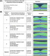
(A) Illustration of the unique features of a normal fovea detectable on optical coherence tomography. (B) Illustration of typical and atypical grades of foveal hypoplasia. All grades of foveal hypoplasia have incursion of inner retinal layers. Atypical (more...)
Cutaneous Manifestations
The skin and hair color of an individual with albinism may change with age (see Table 1). Since evaluating skin hypopigmentation can be subjective and assessment of reduced pigmentation can vary by ethnicity, assessing skin pigmentation in relation to unaffected family members and inquiring about whether the skin tans or burns with sun exposure is helpful. Skin and hair pigmentation scores help to objectively determine effects of hypopigmentation and may guide skin protection recommendations. Note that while these scales are useful, they are not always implemented in busy clinics, where questions are often confined to whether skin tans versus burns.
Table 1.
Skin and Hair Pigmentation Scores
View in own window
| Skin and Hair Pigmentation Scores |
|---|
|
Skin pigmentation score
|
|
Score
|
Color
|
Tanning
|
Other
|
|
1
| White | None | |
|
2
| White | Some | May have pigmented nevi |
|
3
| Pale | Some visible tanning | |
|
4
| Pale | Visible tanning | |
|
5
| Normal | Good tanning | |
|
Hair pigmentation score
|
|
Score
|
Color
|
|
1
| Completely white |
|
2
| Silvery white |
|
3
| White with yellowish touch |
|
4
| Whitish blond |
|
5
| Pale blond |
|
6
| Medium blond |
|
7
| Dark blond |
|
8
| Red, reddish blond |
|
9
| Medium brown |
|
10
| Dark brown, black |
Loss of pigmentation in the skin leads to a variety of psychosocial morbidities and susceptibility to diseases of the skin.
Diseases of the skin. Regardless of their skin tone, all individuals with albinism are at increased risk of skin diseases associated with ultraviolet (UV) light exposure [Ma et al 2023]. The most significant of these is skin cancer, which includes all of the UV light-driven cancers, including squamous cell carcinoma, basal cell carcinoma, melanoma, and less commonly Merkel cell carcinoma [Ma et al 2023]. Individuals with OCA are also at higher risk for sunburns and general sun sensitivity, which can limit outdoor activities where use of sun-protective clothing or sunscreen is not possible. With inappropriate sun exposure, freckles, nevi, and lentigines, which can have cosmetic implications, can develop. Excess UV light exposure can also increase the rate of cutaneous signs of aging [Krutmann et al 2021].
Psychosocial morbidities. The loss of pigmentation tends to have a greater psychosocial effect on individuals with darker skin tone, as the loss of pigmentation is more dramatic [Franklin et al 2018, Marçon & Maia 2019, Anshelevich et al 2021]. In certain African communities, for example, albinism can lead to social isolation [Franklin et al 2018, Marçon & Maia 2019, Nakkazi 2019, Anshelevich et al 2021].
Nomenclature
It is appropriate to classify nonsyndromic OCA according to the gene involved rather than by phenotype (i.e., extent of skin and ophthalmologic involvement). Thus, two former classifications (based on skin and ophthalmologic findings and/or mode of inheritance) are confusing and no longer valid. This is partly due to phenotypic heterogeneity in different races despite having the same genetic variants. The authors recommend that these terms no longer be used.
"Brown" OCA, described initially in Nigeria and Ghana and considered a separate entity based on early family studies, is now known to be part of the phenotypic continuum of
OCA2-related OCA, which encompasses "classic" OCA (yellow/blond hair, creamy-tan skin, and blue/hazel irides) and more pigmented phenotypes like "brown" OCA [
King et al 1985,
Manga et al 2001].
Autosomal recessive ocular albinism (AROA). Individuals with near-normal skin and hair pigmentation, ocular hypopigmentation, and variants in OCA-related genes have been classified as AROA. In a cohort of individuals with suspected AROA, genetic testing revealed a significant proportion of individuals had
hypomorphic TYR variants [
Hutton & Spritz 2008] (see
Table 2). Variants in
OCA2 and
TYRP1 have also been reported to cause AROA [
Hutton & Spritz 2008].
2. Genetic Causes of Oculocutaneous Albinism and Ocular Albinism
Table 2 and Table 3 provide information about the genes that cause oculocutaneous albinism (OCA) and ocular albinism (OA):
Table 2 summarizes genes known to be associated with nonsyndromic OCA and OA. All nonsyndromic OCA is inherited in an
autosomal recessive manner. OA (which is always nonsyndromic) is inherited in an
X-linked manner.
Table 2.
Nonsyndromic Oculocutaneous Albinism and Ocular Albinism by Gene
View in own window
| Gene 1, 2 | Disorder | % of All OCA | Comments |
|---|
|
TYR
| OCA1
(OMIM 606933) | 42% | OCA1 is broadly divided into OCA1A & OCA1B:
|
|
OCA2
| OCA2
(OMIM 611409) | 28% |
|
|
TYRP1
| OCA3
(OMIM 115501) | 2.1% | Phenotype (previously described as "rufous" albinism) is characterized by red-bronze skin color, ginger-red hair, & blue or brown irides. OCA3 is more common in African populations than in other populations (e.g., South Asian, European).
|
SLC45A2
(MATP) |
OCA4
| 11% |
|
|
SLC24A5
| OCA6
(OMIM 609802) | 3% | Ocular features overlap w/other forms of OCA. Typically, skin is hypopigmented w/ability to tan in some persons. Hair color can range from white to brown.
|
LRMDA
(C10orf11) | OCA7
(OMIM 614537) | <1% |
|
DCT
(TYRP2) | OCA8
(OMIM 191275) | <1% | Rare; assoc w/mild hair & skin hypopigmentation |
GPR143
(OA1) | OA1
(OMIM 300808) | 7% | Clinically, only ocular hypopigmentation is present. However, the ocular phenotype of OA & OCA overlap. In heterozygous females (i.e., carriers), "mud-splattered" fundus has been described due to interspersed regions of pigmentation.
|
OA = ocular albinism; OCA = oculocutaneous albinism
- 1.
Where applicable, former gene symbols are listed in parenthesis after the current HGNC-approved gene symbol.
- 2.
Genes are ordered by frequency of causation of OCA [Lasseaux et al 2018]; however, these are based on predominantly European populations. Variants in OCA2 and TYRP1 are more common in sub-Saharan Africa. SLC45A2 variants are more common in Japan.
- 3.
"Brown" OCA, described initially in Nigeria and Ghana and considered a separate entity based on early family studies, is now known to be part of the phenotypic continuum of OCA2-related OCA (see Nomenclature).
- 4.
Table 3.
Syndromic Oculocutaneous Albinism: Genes and Distinguishing Clinical Features
View in own window
| Genes | Syndrome 1 | % of All OCA | Ocular & Cutaneous Features | Other Clinical Features |
|---|
AP3B1
AP3D1
BLOC1S3
BLOC1S5
BLOC1S6
DTNBP1
HPS1
HPS3
HPS4
HPS5
HPS6
| Hermansky-Pudlak syndrome (HPS) | 4% | Nearly all children w/HPS-related albinism have infantile nystagmus. Hair color ranges from white to brown. Skin color is generally at least a shade lighter than that of other family members. Only grades 3 & 4 foveal hypoplasia have been observed w/reduced visual acuity.
|
|
|
LYST
|
Chediak-Higashi syndrome
| <1% | OCA features are less severe & are hence referred to as "partial" OCA. | Immunodeficiency & bleeding diathesis |
MLPH
MYO5A
RAB27A
| Griscelli syndrome(GS) (OMIM PS214450) | <1% |
| The 3 subtypes are:
GS1: + neurologic deficits but no immunologic dysfunction GS2: ± neurologic deficits; + immunologic dysfunction GS3: no neurologic or immunologic deficits
|
Waardenburg syndrome is an auditory-pigmentary syndrome with sensorineural hearing loss, neurologic deficits in some affected individuals, and variable hypopigmentation of hair, skin, and eyes. It is debated whether Waardenburg syndrome should be classified as a syndromic form of OCA, since all characteristic ocular features seen in OCA may not be present. See Waardenburg Syndrome Type I.
4. Evaluation Strategies to Identify the Genetic Cause of Oculocutaneous Albinism or Ocular Albinism in a Proband
Establishing a specific genetic cause of oculocutaneous albinism (OCA) or ocular albinism (OA) in a proband:
Can aid in discussions of prognosis (which are beyond the scope of this
GeneReview) and
genetic counseling;
Usually involves a medical history, physical examination, laboratory testing, family history, and
genomic/genetic testing;
Can influence treatments and surveillance of disease, particularly in
syndromic forms of OCA due to implications to systemic health.
Medical History
Individuals with OCA often present with infantile nystagmus, poor vision, and hypopigmentation (see Clinical Characteristics). Photophobia may be present.
Identifying manifestations suggestive of syndromic forms of OCA is important, as management differs between nonsyndromic OCA (see Table 2) and syndromic forms of OCA (see Table 3). Syndromic OCA is suggested when findings such as the following are present:
Immune dysfunction
Bleeding diathesis, including easy bruising, epistaxis, and prolonged bleeding after minor procedures or surgery
Neurologic deficits such as developmental delay / intellectual disability and seizures
Pulmonary fibrosis and granulomatous colitis
Physical Examination and Clinic-Based Investigations
Detailed ophthalmologic evaluation should include refraction, best corrected visual acuity, ocular motility (strabismus and nystagmus characteristics), measurement of anomalous head posture, slit lamp examination and/or optical coherence tomography (OCT) to detect iris transillumination defects (TIDs), assessment of fundus hypopigmentation, characterization of foveal morphology using OCT, and optic nerve misrouting using visual evoked potentials (VEPs) (see Clinical Characteristics, Eyes/Vision).
Examining parents of children with OCA can also be helpful, as they may exhibit subclinical features such as low grades of iris TIDs, fundus hypopigmentation, and foveal hypoplasia [Kuht et al 2022b]. This is particularly useful when there is diagnostic uncertainty and/or limited phenotype information can be obtained from an uncooperative young child presenting with infantile nystagmus.
Similarly, unaffected female carriers of GPR143 variants / X-linked OA can have iris TIDs, a "mud-splattered" fundus appearance, and foveal hypoplasia [Charles et al 1993, Khan et al 2018].
Due to the shared ocular and cutaneous phenotypic characteristics, differentiating between syndromic and nonsyndromic OCA can be challenging (see Clinical Characteristics).
Family History
A three-generation family history should be taken, with attention to relatives with manifestations of OCA or OA. Note that relevant findings can be documented through direct examination and/or review of medical records, including results of molecular genetic testing. A family history consistent with X-linked inheritance (e.g., no male-to-male transmission) may be helpful in differentiating nonsyndromic GPR143-related OA (an X-linked disorder) from OCA.
Molecular Genetic Testing
Molecular genetic testing approaches can include a combination of targeted testing (multigene panel and chromosomal microarray to detect recurrent deletions) and comprehensive genomic testing (exome sequencing or genome sequencing). Gene-targeted testing (see Option 1) requires the clinician to hypothesize which gene(s) are likely involved, whereas genomic testing (see Option 2) does not.
Option 1
A multigene panel that includes some or all of the genes listed in Tables 2 and 3 is most likely to identify the genetic cause of the condition while limiting identification of variants of uncertain significance and pathogenic variants in genes that do not explain the underlying phenotype. Notes: (1) The genes included in the panel and the diagnostic sensitivity of the testing used for each gene vary by laboratory and are likely to change over time. (2) Some multigene panels may include genes not associated with the condition discussed in this GeneReview. Of note, given the rarity of some of the genes associated with OCA, some panels may not include all the genes mentioned in this overview. (3) In some laboratories, panel options may include a custom laboratory-designed panel and/or custom phenotype-focused exome analysis that includes genes specified by the clinician. (4) Methods used in a panel may include sequence analysis, deletion/duplication analysis, and/or other non-sequencing-based tests.
For an introduction to multigene panels click here. More detailed information for clinicians ordering genetic tests can be found here.
Chromosomal microarray analysis (CMA) using oligonucleotide or SNP arrays to detect large deletions/duplications that cannot be detected by sequence analysis should be considered in OCA, as several large recurrent deletions have been identified.
Note: (1) The most common OCA2 pathogenic variant in African and African American populations is a recurrent 2.7-kb deletion spanning exon 7 of the gene. The 2.7-kb deletion is less common in the US African American population, but has been identified in the Puerto Rican population. (2) Other larger deletions including OCA2 have been reported [Kedda et al 1994, Spritz et al 1995, Stevens et al 1995, Durham-Pierre et al 1996, Puri et al 1997, Stevens et al 1997, Kerr et al 2000, Santiago Borrero et al 2006].
For an introduction to CMA click here. More detailed information for clinicians ordering genetic tests can be found here.
Option 2
Comprehensive
genomic testing (which does not require the clinician to determine which gene[s] are likely involved) may be considered. Exome sequencing is most commonly used; genome sequencing is also possible.
For an introduction to comprehensive genomic testing click here. More detailed information for clinicians ordering genomic testing can be found here.
5. Genetic Counseling
Genetic counseling is the process of providing individuals and families with
information on the nature, mode(s) of inheritance, and implications of genetic disorders to help them
make informed medical and personal decisions. The following section deals with genetic
risk assessment and the use of family history and genetic testing to clarify genetic
status for family members; it is not meant to address all personal, cultural, or
ethical issues that may arise or to substitute for consultation with a genetics
professional. —ED.
Autosomal Recessive Inheritance – Risk to Family Members
Parents of a proband
Sibs of a proband
Offspring of a proband. The offspring of an individual with autosomal recessive OCA are obligate heterozygotes (carriers) for an OCA-causing pathogenic variant.
Other family members. Each sib of the proband's parents is at a 50% risk of being a carrier of an OCA-causing pathogenic variant.
Carrier detection. Carrier testing for at-risk relatives requires prior identification of the OCA-causing pathogenic variants in the family.
X-Linked Inheritance – Risk to Family Members
Parents of a male proband
The father of an affected male will not have the disorder nor will he be
hemizygous for the
GPR143 pathogenic variant; therefore, he does not require further evaluation/testing.
Molecular genetic testing of the mother is recommended to confirm her genetic status and to allow reliable
recurrence risk assessment.
Sibs of a male proband. The risk to sibs depends on the genetic status of the mother:
Females who inherit the
pathogenic variant will be heterozygotes (i.e., carriers). Heterozygotes are usually not affected, although they can exhibit iris transillumination defects, a "mud-splattered" fundus appearance, and foveal hypoplasia [
Charles et al 1993,
Khan et al 2018].
If the
proband represents a
simplex case and if the
pathogenic variant cannot be detected in the leukocyte DNA of the mother, the risk to sibs is presumed to be low but greater than that of the general population because of the possibility of maternal
germline mosaicism.
Offspring of a male proband. Affected males transmit the GPR143 pathogenic variant to all of their daughters and none of their sons.
Other family members. The maternal aunts and maternal cousins of a male proband may be at risk of having a GPR143 pathogenic variant.
Note: Molecular genetic testing may be able to identify the family member in whom a de novo pathogenic variant arose, information that could help determine genetic risk status of the extended family.
Heterozygote detection. Identification of female heterozygotes requires prior identification of the GPR143 pathogenic variants in the family.
Note: Females who are heterozygotes (carriers) for this X-linked disorder will be heterozygotes and will usually not be affected, although they can exhibit iris transillumination defects, a "mud-splattered" fundus appearance, and foveal hypoplasia [Charles et al 1993, Khan et al 2018].
Prenatal Testing and Preimplantation Genetic Testing
Once the OCA- or OA-causing pathogenic variant(s) have been identified in an affected family member, prenatal and preimplantation genetic testing are possible.
Differences in perspective may exist among medical professionals and within families regarding the use of prenatal testing. While most centers would consider use of prenatal testing to be a personal decision, discussion of these issues may be helpful.
6. Management
At present, no curative treatments are available for albinism.
Supportive treatments are aimed at optimizing vision, managing clinical manifestations (e.g., nystagmus), and reducing risks of complications of cutaneous albinism (e.g., skin cancer).
Eyes/Vision
Refractive errors and optimizing visual acuity. Correction of refractive errors found in most individuals with albinism (either hyperopia or myopia and astigmatism) with spectacles or (when age appropriate) contact lenses can optimize visual acuity.
Because anisometropic and ametropic amblyopia may develop in children with albinism, prompt correction of refractive errors in children to reduce risk for amblyopia is essential. If amblyopia is present following a refractive adaptation period of 16-18 weeks, occlusion therapy should commence.
Optical coherence tomography is useful in characterizing retinal development based on foveal hypoplasia grades (see ) [Thomas et al 2011b]. If there is a mismatch between the measured visual acuity and expected visual potential (based on the grade of foveal hypoplasia), other causes of reduced vision should be investigated and, if identified, treated accordingly.
Low vision aids may be useful in some individuals with albinism; these are typically issued following full refractive correction. Magnification apparatuses such as telescopes and near magnifiers may be used in individuals with significantly reduced visual acuity [Liu et al 2021].
Management of nystagmus, strabismus, and anomalous head posture (AHP). Vision of individuals with albinism can be improved by treatments that aim to reduce the intensity of the nystagmus, lengthen the foveation periods in the null zone (i.e., the brief period of time during nystagmus when the eyes are relatively still and visual information is captured by the fovea), or correct AHP by moving the null zone into primary position.
AHP can have a significant effect on quality of life, causing neck issues, headache, and cosmetic issues. When AHP prevents children with low visual acuity from looking through their glasses, they are at risk of developing amblyopia. The Anderson-Kestenbaum procedure, the standard surgical approach for AHP, is generally performed after age two years.
Infantile nystagmus. In studying the effect of gabapentin or memantine on visual function in 16 individuals with infantile nystagmus,
McLean et al [2007] reported significantly improved visual acuity in individuals with
idiopathic infantile nystagmus; however, the change in those with a secondary cause of nystagmus (e.g., albinism) was not statistically significant. Nonetheless, nystagmus intensity and foveation improved on eye movement recordings after medication in all individuals.
The evidence for the use of contact lenses to improve characteristics of nystagmus is unclear.
Reducing glare. Dark glasses or transition lenses may be helpful, but many individuals with albinism prefer to go without the tint because of the reduction in vision from the dark lenses.
A hat with a brim may be helpful to reduce overhead glare and, importantly, provide some sun protection for the face.
Other approaches to alleviate glare described in the literature with either limited success or complications arising from the intervention include surgical implants or various designs of modified contact lenses. Because the evidence level is restricted to case reports, these interventions are not recommended.
Skin
Individuals with cutaneous albinism are encouraged to have complete skin examinations at least yearly to identify evidence of sun damage or early signs of skin cancer. At these visits, education regarding methods of sun protection is stressed, including the following:
In most circumstances, especially when the parents do not have albinism, children with albinism are referred to a pediatric dermatologist at a young age to ensure that parents and family members are properly educated regarding sun protection.
It is problematic for individuals with albinism to self-monitor their skin. Whereas skin cancers such as squamous cell carcinoma and basal cell carcinoma are clinically the same in individuals with normal pigmentation and in individuals with albinism, melanoma is different.
Melanoma, the most dangerous of all skin cancers, is clinically diagnosed based on changes in pigmentation patterns [Tsao et al 2015]. In individuals with normal skin pigmentation, clinicians and the individuals themselves monitor pigmented lesions for changes in symmetry, color, border, and lesion diameter to identify those suspicious for melanoma that require a biopsy for pathologic confirmation. In contrast, in individuals with albinism, melanomas lack pigment, making traditional methods for early diagnosis very difficult. For these reasons, yearly total body skin examinations by a trained dermatologist are highly recommended. A low threshold for skin biopsy is important. Treatment of skin cancer in individuals with albinism is the same as the general population.
Educational Management
The following information represents typical management recommendations for children of school age with educational issues related to decreased visual acuity.
Ages 0-3 years. Referral to an early intervention program is recommended for access to special educators and sensory impairment specialists. In the US, early intervention is a federally funded program available in all states that provides in-home services to target individual therapy needs.
Ages 3-5 years. In the US, assessment of low vision needs and developmental assessment is recommended before entering school. Before placement, an evaluation is made to determine needed services and therapies and an individualized education plan (IEP) is developed for those who qualify based on established motor, language, social, or cognitive delay. The early intervention program typically assists with this transition. Developmental preschool is center based; for children too medically unstable to attend, home-based services are provided.
All ages. Consultation with a developmental pediatrician is recommended to ensure the involvement of appropriate community, state, and educational agencies (US) and to support parents in maximizing quality of life. Some issues to consider:
IEP services:
An IEP provides specially designed instruction and related services to children who qualify.
IEP services will be reviewed annually to determine whether any changes are needed.
Special education law requires that children participating in an IEP be in the least restrictive environment feasible at school and included in general education as much as possible, when and where appropriate.
Vision and hearing consultants should be a part of the child's IEP team to support access to academic material.
PT, OT, and speech services will be provided in the IEP to the extent that the need affects the child's access to academic material. Beyond that, private supportive therapies based on the affected individual's needs may be considered. Specific recommendations regarding type of therapy can be made by a developmental pediatrician.
As a child enters the teen years, a transition plan should be discussed and incorporated in the IEP. For those receiving IEP services, the public school district is required to provide services until age 21.
A 504 plan (Section 504: a US federal statute that prohibits discrimination based on disability) can be considered for those who require accommodations or modifications such as front-of-class seating, assistive technology devices, use of magnifiers, classroom scribes, extra time between classes, modified assignments, and enlarged text.
Developmental Disabilities Administration (DDA) enrollment is recommended. DDA is a US public agency that provides services and support to qualified individuals. Eligibility differs by state but is typically determined by diagnosis and/or associated cognitive/adaptive disabilities.
Families with limited income and resources may also qualify for supplemental security income (SSI) for their child with a disability.
Surveillance
Eyes/vison. For children younger than age 16 years, the following evaluations are recommended annually:
Ophthalmologic examination (including assessment of refractive errors; strabismus and/or anomalous head posture; and the need for filter glasses)
Assessment of psychosocial needs and support
Assessment of educational needs and support
For adults, the following evaluations are recommended:
Skin. An annual to biennial skin examination is recommended to assess for evidence of sun-related skin damage and/or precancerous or cancerous lesions, especially in areas of high-intensity or prolonged sunlight exposure.
Agents/Circumstances to Avoid
Prolonged unprotected sun exposure should be avoided.
Chapter Notes
Acknowledgments
The authors thank the patients, families, and support groups that volunteer their time and engage in research to further our understanding of albinism. The authors thank their research teams, including the Ulverscroft Eye Unit (University of Leicester, UK), Zippin Lab (Weill Cornell Medical College of Cornell University, USA), and the National Eye Institute (NIH, USA).
MT gratefully acknowledges the support from the National Institute for Health Research (CL-2017-11-003), Medical Research Council, Wellcome Trust, Fight for Sight, Ulverscroft Foundation, and the Academy of Medical Sciences.
JZ gratefully acknowledges the support from the National Institute of Arthritis, Musculoskeletal and Skin Diseases (R01-AR077664).
BB gratefully acknowledges the support from the Intramural Program at the National Eye Institute, National Institutes of Health.