U.S. flag

An official website of the United States government

NCBI Bookshelf. A service of the National Library of Medicine, National Institutes of Health.

Adam MP, Feldman J, Mirzaa GM, et al., editors. GeneReviews® [Internet]. Seattle (WA): University of Washington, Seattle; 1993-2024.

Cover of GeneReviews®

GeneReviews® [Internet].

Show details

PLA2G6-Associated Neurodegeneration

Synonyms: NBIA2, PLA2G6-Related Disorders, PLAN

, MS, CGC, , MA, MRCPCH, PhD, , MD, FRCP, FMedSci, , MD, and , MD.

Author Information and Affiliations

Initial Posting: ; Last Update: March 23, 2017.

Estimated reading time: 24 minutes

Summary

Clinical characteristics.

PLA2G6-associated neurodegeneration (PLAN) comprises a continuum of three phenotypes with overlapping clinical and radiologic features:

  • Infantile neuroaxonal dystrophy (INAD)
  • Atypical neuroaxonal dystrophy (atypical NAD)
  • PLA2G6-related dystonia-parkinsonism

INAD usually begins between ages six months and three years with psychomotor regression or delay, hypotonia, and progressive spastic tetraparesis. Many affected children never learn to walk or lose the ability shortly after attaining it. Strabismus, nystagmus, and optic atrophy are common. Disease progression is rapid, resulting in severe spasticity, progressive cognitive decline, and visual impairment. Many affected children do not survive beyond their first decade.

Atypical NAD shows more phenotypic variability than INAD. In general, onset is in early childhood but can be as late as the end of the second decade. The presenting signs may be gait instability, ataxia, or speech delay and autistic features, which are sometimes the only evidence of disease for a year or more. Strabismus, nystagmus, and optic atrophy are common. Neuropsychiatric disturbances including impulsivity, poor attention span, hyperactivity, and emotional lability are also common. The course is fairly stable during early childhood and resembles static encephalopathy but is followed by neurologic deterioration between ages seven and 12 years.

PLA2G6-related dystonia-parkinsonism has a variable age of onset, but most individuals present in early adulthood with gait disturbance or neuropsychiatric changes. Affected individuals consistently develop dystonia and parkinsonism (which may be accompanied by rapid cognitive decline) in their late teens to early twenties. Dystonia is most common in the hands and feet but may be more generalized. The most common features of parkinsonism in these individuals are bradykinesia, resting tremor, rigidity, and postural instability.

Diagnosis/testing.

The diagnosis of PLA2G6-associated neurodegeneration is established in a proband by identification of biallelic pathogenic variants in PLA2G6 on molecular genetic testing. The diagnosis of INAD or atypical NAD can be established in a proband with no identified PLA2G6 pathogenic variants by electron microscopic examination of nerve biopsies for dystrophic axons (axonal spheroids).

Management.

Treatment of manifestations:

  • Individuals with INAD and atypical NAD. Routine pharmacologic treatment of spasticity and seizures; trial of oral or intrathecal baclofen for dystonia associated with atypical INAD; treatment by a psychiatrist for those with later-onset neuropsychiatric symptoms; fiber supplements and/or stool softener treatment for constipation; control of secretions with transdermal scopolamine patch as needed; feeding modifications as needed to prevent aspiration pneumonia and achieve adequate nutrition.
  • Individuals with PLA2G6-related dystonia-parkinsonism. Consider treatment with dopaminergic agents; treatment of neuropsychiatric symptoms by a psychiatrist; evaluation by physical therapy for management of postural instability and gait difficulties; occupational therapy to assist with activities of daily living; feeding modifications as needed to prevent aspiration pneumonia and achieve adequate nutrition.

Prevention of secondary complications: Early physical therapy and orthopedic management to prevent contractures as the disease progresses; body temperature monitors may be required for individuals with progressive autonomic involvement to identify dangerous fluctuations in core body temperature.

Surveillance: Periodic assessment of vision and hearing of nonverbal children is indicated as needed to determine the level of sensory deficits.

Genetic counseling.

PLA2G6-associated neurodegeneration is inherited in an autosomal recessive manner. At conception, each sib of an affected individual has a 25% chance of being affected, a 50% chance of being an asymptomatic carrier, and a 25% chance of being unaffected and not a carrier. Carrier testing for at-risk family members, prenatal testing for a pregnancy at increased risk, and preimplantation genetic testing are possible if the pathogenic variants in the family are known.

GeneReview Scope

PLA2G6-Associated Neurodegeneration: Included Phenotypes 1
  • Infantile neuroaxonal dystrophy (INAD)
  • Atypical neuroaxonal dystrophy (atypical NAD)
  • PLA2G6-related dystonia-parkinsonism

For synonyms and outdated names see Nomenclature.

1.

For other genetic causes of these phenotypes see Differential Diagnosis.

Diagnosis

Suggestive Findings

PLA2G6-associated neurodegeneration (PLAN) comprises a continuum of three phenotypes with overlapping clinical and radiologic features:

  • Infantile neuroaxonal dystrophy (INAD)
  • Atypical neuroaxonal dystrophy (atypical NAD)
  • PLA2G6-related dystonia-parkinsonism

Infantile Neuroaxonal Dystrophy (INAD)

PLA2G6-associated INAD should be suspected in individuals with the following clinical, laboratory, radiographic, and neurophysiologic features.

Clinical

  • Onset before age three years
  • Psychomotor regression (most common presenting feature)
  • Early truncal hypotonia followed by spastic tetraparesis (usually with hyperreflexia in the early disease stages with progression to areflexia later in the disease course)
  • Visual abnormalities: strabismus, nystagmus, optic atrophy

Laboratory

  • Elevated aspartate aminotransferase / alanine aminotransferase ratio
  • Elevated lactate dehydrogenase

Radiographic

  • Cerebellar atrophy (See Figure 1.)
  • T2-weighted MRI of the brain: hypointense globus pallidus (indicating iron accumulation), cortical cerebellar hyperintensities consistent with cerebellar gliosis, white matter abnormalities, thin vertically oriented corpus callosum (see Figure 1), and hypertrophy of the clava [Illingworth et al 2014, Al-Maawali et al 2016]
Figure 1. A.

Figure 1

A. Left axial image shows high brain iron in the globus pallidus (see arrow) on T2-weighted MRI. B. Right sagittal image shows cerebellar atrophy (see arrow).

Neurophysiologic

  • EMG (electromyogram). Evidence of denervation
  • EEG (electroencephalogram). Fast rhythms
  • VEP (visual evoked potential). Delayed with reduced amplitudes
  • NCV (nerve conduction velocity). Distal axonal-type sensorimotor neuropathy
  • Seizures that may present early or late in the disease course [Wu et al 2009]

Atypical Neuroaxonal Dystrophy (NAD)

PLA2G6-associated atypical NAD) should be suspected in individuals with the following clinical, radiographic, and neurophysiologic features.

Clinical

  • Onset before age 20 years
  • Psychomotor regression
  • Gait abnormalities
  • Prominent expressive language difficulties
  • Psychiatric/behavioral abnormalities including autistic-like behavior
  • Visual abnormalities: nystagmus, optic atrophy
  • Spasticity (without preceding hypotonia)
  • Joint contractures
  • Progressive dystonia and dysarthria
  • Disease progression slower than in INAD

Radiographic

  • Cerebellar atrophy
  • T2-weighted MRI of the brain: hypointense globus pallidus (indicating iron accumulation)

Neurophysiologic

  • VEP. Delayed with reduced amplitudes
  • Seizures

PLA2G6-Related Dystonia-Parkinsonism

PLA2G6-related dystonia-parkinsonism should be suspected in individuals with the following clinical and radiographic features.

Clinical

  • Onset varying from childhood to young adulthood
  • Parkinsonism (tremor, bradykinesia, rigidity, and markedly impaired postural responses)
  • Dystonia
  • Dysarthria
  • Autonomic involvement (e.g., cold/blue hands and feet, difficulty regulating core body temperature, constipation)
  • Cognitive decline
  • Neuropsychiatric changes
  • Initial dramatic response to dopaminergic treatment followed by the early development of dyskinesias

Radiographic

  • Cerebral atrophy
  • Cerebellar atrophy
  • Abnormal brain iron accumulation in the globus pallidus, substantia nigra, and/or striatum; findings are variable and may not be evident on MRI studies until late in the disease course for some individuals.
  • Reduced dopamine transporter labeling similar to that seen in idiopathic Parkinson disease
  • In some individuals, frontotemporal atrophy/hypoperfusion on single-photon emission computed tomography

Establishing the Diagnosis

The diagnosis of PLA2G6-associated neurodegeneration (PLAN) is established in a proband by identification of biallelic pathogenic variants in PLA2G6 on molecular genetic testing (see Table 1).

Molecular genetic testing approaches can include single-gene testing, use of a multigene panel, and more comprehensive genomic testing:

  • Single-gene testing. Sequence analysis of PLA2G6 is performed first and followed by gene-targeted deletion/duplication analysis if only one or no pathogenic variant is found.
  • A multigene panel that includes PLA2G6 and other genes of interest (see Differential Diagnosis) may also be considered. Note: (1) The genes included in the panel and the diagnostic sensitivity of the testing used for each gene vary by laboratory and are likely to change over time. (2) Some multigene panels may include genes not associated with the condition discussed in this GeneReview; thus, clinicians need to determine which multigene panel is most likely to identify the genetic cause of the condition while limiting identification of variants of uncertain significance and pathogenic variants in genes that do not explain the underlying phenotype. (3) Methods used in a panel may include sequence analysis, deletion/duplication analysis, and/or other non-sequencing-based tests.
    For an introduction to multigene panels click here. More detailed information for clinicians ordering genetic tests can be found here.
  • More comprehensive genomic testing (when available) including exome sequencing and genome sequencing may be considered. Such testing may provide or suggest a diagnosis not previously considered (e.g., mutation of a different gene or genes that results in a similar clinical presentation).
    For an introduction to comprehensive genomic testing click here. More detailed information for clinicians ordering genomic testing can be found here.

Table 1.

Molecular Genetic Testing Used in PLA2G6-Associated Neurodegeneration

Gene 1MethodProportion of Probands with Pathogenic Variants 2 Detectable by Method
PLA2G6 Sequence analysis 3~85% 4
Gene-targeted deletion/duplication analysis 5≤12.5% 6
Unknown 7NA
1.
2.

See Molecular Genetics for information on allelic variants detected in this gene.

3.

Sequence analysis detects variants that are benign, likely benign, of uncertain significance, likely pathogenic, or pathogenic. Variants may include small intragenic deletions/insertions and missense, nonsense, and splice site variants; typically, exon or whole-gene deletions/duplications are not detected. For issues to consider in interpretation of sequence analysis results, click here.

4.

Of all individuals identified with PLA2G6 pathogenic variants, approximately 10% have only one pathogenic variant identified [NBIA International Mutation Database, unpublished data].

5.

Gene-targeted deletion/duplication analysis detects intragenic deletions or duplications. Methods used may include a range of techniques such as quantitative PCR, long-range PCR, multiplex ligation-dependent probe amplification (MLPA), and a gene-targeted microarray designed to detect single-exon deletions or duplications.

6.

Deletion and duplication of multiple exons have been identified Crompton et al [2010] (see Molecular Genetics, Pathogenic variants).

7.

Linkage data support the presence of at least one additional INAD locus [Morgan et al 2006].

Tissue biopsy. If no PLA2G6 pathogenic variants are identified but the evolving phenotype remains most consistent with INAD or atypical NAD, a biopsy for identification of dystrophic axons (axonal spheroids) can be considered. Electron microscopic (EM) examination of nerve ultrastructure should be done on one of the following preferred tissues: conjunctiva, skin, rectum, muscle, or other peripheral nerve (sural). Histopathologic evidence of dystrophic axons on biopsy from one or more of the following tissues, viewed by EM, includes:

  • Membranotubular profiles;
  • Mitochondrial aggregates;
  • Increased axonal diameter and thinned membrane.

Note: (1) Because axonal spheroids accumulate with age and may not be evident in all tissues, individuals suspected to have INAD or atypical NAD without identifiable PLA2G6 pathogenic variants may require multiple biopsies over time before axonal spheroids are identified. (2) Peripheral spheroids have not been described in pathologic specimens from persons with PLA2G6-associated dystonia-parkinsonism; however, limited pathologic material has been available thus far from this group.

Clinical Characteristics

Clinical Description

Infantile neuroaxonal dystrophy (INAD). Onset of INAD usually occurs between ages six months and three years. The disease presents with psychomotor regression (i.e., loss of previously acquired milestones) or delay, delayed walking, or gait disturbance. A single individual with neonatal onset has been reported, with severe hypotonia and marked weakness [Fusco et al 2015].

Truncal hypotonia is observed early in the disease course. Over time, affected persons develop a spastic tetraparesis, with symmetric pyramidal tract signs on clinical examination.

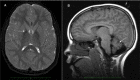
Visual signs and symptoms are common. Strabismus and nystagmus are early features of the disease. Later optic atrophy occurs in most individuals. Optic atrophy may be observed early as optic nerve pallor; thin optic chiasm and tracts have also been reported on brain MRI [Farina et al 1999].

Seizures occur in a minority of individuals as a later symptom [Nardocci et al 1999, Wu et al 2009].

Autonomic involvement may present early as constipation or cold extremities. With progression, some individuals require body temperature monitors because of dangerous fluctuations in core body temperature.

The progression of disease is usually rapid. Many affected children never learn to walk or lose this ability shortly after attaining it. During the end stages of disease, severe spasticity, progressive cognitive decline, and visual impairment result in a vegetative state. Death occurs as a result of secondary illnesses such as aspiration pneumonia, associated with bulbar dysfunction. Many affected children do not survive beyond their first decade, but some survive into their teens or later. Supportive care can contribute to a longer life span by reducing the risk of infection and other complications.

Atypical NAD. Whereas the features of INAD are relatively homogeneous, atypical disease is quite varied.

In general, onset in atypical NAD is in early childhood but can be as late as the late teens. In a series of 13 individuals, four had onset by age three years but a fairly stable course during early childhood resembling static encephalopathy, followed by neurologic deterioration between ages seven and 12 years [Nardocci et al 1999].

The presenting signs and symptoms may be similar to INAD, including gait instability or ataxia. Others may present with speech delay and autistic features, which may remain as the only evidence of disease for a year or more, given the slow progression of atypical NAD compared to INAD [Gregory et al 2008].

Although spastic tetraparesis is evident late in the disease, it is rarely preceded by early truncal hypotonia. In contrast to classic disease, extrapyramidal findings (i.e., dystonia and dysarthria) predominate in atypical NAD. Eye findings are similar to those seen in classic INAD. Neuropsychiatric disturbances including impulsivity, poor attention span, hyperactivity, and emotional lability are also common [Gregory et al 2008].

Atypical NAD is rare, and the life span is not known; however, it is expected to be longer than that observed in classic disease.

PLA2G6-related dystonia-parkinsonism. To date, only a small number of affected individuals have been described [Karkheiran et al 2015]. Age at onset has varied from four to 37 years [Paisán-Ruiz et al 2009, Paisán-Ruiz et al 2010, Yoshino et al 2010, Bower et al 2011, Paisán-Ruiz et al 2012, Virmani et al 2014]; however, the majority have presented in early adulthood (late teens to 20s). Of those with childhood onset, one presented with foot drag and dystonia at age ten years and the other two children presented with an unsteady gait at ages six and eight years. The youngest individual presented with stuttering speech, clumsiness, and dyslexia at age four years – findings that may not be related to the PLA2G6-associated neurodegeneration (PLAN). In young adults, initial symptoms are frequently neuropsychiatric, including depression, personality changes, aggression, delusions, or paranoia. Gait disturbance is also common at presentation.

Regardless of the age at onset, affected individuals consistently develop dystonia and parkinsonism (which may be accompanied by rapid cognitive decline) in their late teens to early twenties. Neuropsychiatric changes may precede the movement disorder or occur concomitantly. Dystonia is most common in the hands and feet but may be more generalized. The most common features of parkinsonism in these individuals are bradykinesia, resting tremor, rigidity, and postural instability. Of note, it is common to have an initially dramatic positive response to dopaminergic agents; however, this tends to be short-lived and followed quickly by the development of motor fluctuations and dyskinesias.

Neuropathology. Paisán-Ruiz et al [2012] described the neuropathologic findings in seven individuals who spanned the three forms of PLAN. Numerous axonal swellings in the basal ganglia and brain stem were observed in individuals with infantile-onset and adult-onset PLAN. They were also found in the spinal cord in the two individuals for whom cord tissue was available. Lewy bodies were widespread in both those with adult-onset and those with infantile-onset PLAN. In two affected individuals, one with onset at 18 years and the other only specified as "childhood," the Lewy body pathology was comparable to that seen in severe, end-stage Parkinson disease. Tau pathology, to varying degrees, was also found across the PLAN spectrum.

Genotype-Phenotype Correlations

Genotype correlates with phenotype to a limited extent:

  • All individuals with two null alleles of PLA2G6 have INAD.
  • The less severe atypical NAD phenotype is caused almost exclusively by pathogenic missense variants.
  • Common pathogenic variants associated with INAD impair the catalytic activity of the PLA2G6 protein, whereas three pathogenic variants associated with PLA2G6-related dystonia-parkinsonism did not [Engel et al 2010].

Nomenclature

Outdated terms

  • Seitelberger [1952] first described this condition, which was originally named Seitelberger disease.
  • Karak syndrome was described in two sibs with early-onset cerebellar ataxia, dystonia, spasticity, and intellectual decline. Brain MRI findings included cerebellar atrophy and iron accumulation in the globus pallidus and substantia nigra [Mubaidin et al 2003]. Morgan et al [2006] identified pathogenic variants in PLA2G6 in individuals with Karak syndrome, which is now included in the phenotypic spectrum of PLAN and no longer considered a clinically distinct entity; what had been described as Karak syndrome is now referred to as atypical NAD.

Current nomenclature. In addition to INAD, later-onset variants have been called late-infantile, juvenile, or atypical neuroaxonal dystrophy and neurodegeneration with brain iron accumulation (NBIA).

The authors propose the following usage:

  • INAD for early-onset, rapidly progressive disease
  • Atypical NAD for later childhood-onset disease with slower progression and predominant extrapyramidal findings (dystonia, dysarthria). The atypical NAD phenotype is expected to include a broad range of presentations including Karak syndrome.
  • PLA2G6-related dystonia-parkinsonism for adult-onset dystonia-parkinsonism accompanied by cognitive decline and neuropsychiatric changes

Prevalence

Disease prevalence is not established; it is estimated at 1:1,000,000.

Differential Diagnosis

Infantile Neuroaxonal Dystrophy (INAD)

Early diagnosis is challenging because the initial symptoms of psychomotor regression and progression are also observed in other conditions. The observation of an elevated aspartate aminotransferase / alanine aminotransferase ratio and elevated lactate dehydrogenase in combination with these findings is more suspicious for INAD [Kraoua et al 2016].

The degree of weakness early in the disease course may initially direct the clinician toward a myopathy or spinal muscular atrophy.

Cerebellar atrophy can be detected by brain MRI before age two years in some children [Farina et al 1999]. The differential diagnosis for childhood cerebellar atrophy includes infantile neuronal ceroid-lipofuscinosis (CLN1 disease, Santavuori-Haltia), ataxia-telangiectasia, KIF1A-associated hereditary spastic paraplegia (OMIM 610357; see also Hereditary Spastic Paraplegia Overview), and hereditary ataxia; however, cerebellar atrophy usually presents later in individuals with these disorders.

An estimated 40%-50% of individuals with INAD have abnormal iron accumulation in the basal ganglia (primarily the globus pallidus), which is best detected on T2-weighted MRI. For this reason, conditions included in the neurodegeneration with brain iron accumulation (NBIA) category should also be considered in the differential diagnosis of INAD. Individuals with INAD have not been found to have an eye-of-the-tiger sign, which correlates very highly with pantothenate kinase-associated neurodegeneration (PKAN) [Hayflick et al 2003].

Since the identification of PLA2G6 pathogenic variants as causative of INAD, the need for invasive nerve biopsy to aid in diagnosis has decreased. While the presence of axonal spheroids in peripheral tissues remains specific to INAD, spheroids are found in the brain in a few other conditions, including PKAN, idiopathic NBIA, infantile GM2 gangliosidosis (see Hexosaminidase A Deficiency), Niemann-Pick disease type C, and Menkes disease (see ATP7A-Related Copper Transport Disorders).

Atypical Neuroaxonal Dystrophy (NAD)

Initial speech delay and limited social interaction may be consistent with autism.

Spasticity, dystonia, and dysarthria – findings similar to those of other forms of NBIA – eventually predominate; high brain iron in the globus pallidus and substantia nigra has been observed in nearly all individuals, although ascertainment is likely to be biased [Gregory et al 2008]. Therefore, idiopathic NBIA should also be considered in the differential diagnosis of atypical NAD. PKAN may present with similar features.

PLA2G6-Related Dystonia-Parkinsonism

When high brain iron is present and pathogenic variants in PLA2G6 have not been identified, other forms of NBIA should be considered in the differential diagnosis. Atypical PKAN, Kufor-Rakeb syndrome (OMIM 606693), MPAN (mitochondrial membrane protein-associated neurodegeneration), and BPAN (beta-propeller protein-associated neurodegeneration) can present with neuropsychiatric changes, parkinsonism, and dystonia in late childhood or early adulthood. As in PLA2G6-related dystonia-parkinsonism, individuals with MPAN, BPAN, and Kufor-Rakeb syndrome also exhibit cognitive decline.

Other forms of early-onset dystonia-parkinsonism must also be considered, including: dopa-responsive dystonia; Wilson disease; Parkinson disease 2 (PARK2); PARK6, PARK7, and PARK15 (see Parkinson Disease Overview); SLC6A3-related dystonia-parkinsonism; X-linked dystonia-parkinsonism (DYT-TAF1); DYT-ATP1A3; DYT16; and spastic paraplegia 11 (SPG11) [Schneider & Bhatia 2010].

Management

Evaluations Following Initial Diagnosis

To establish the extent of disease and needs in an individual diagnosed with PLA2G6-associated neurodegeneration (PLAN), the following evaluations are recommended if they have not already been completed:

  • Thorough ophthalmologic examination to assess for optic atrophy
  • EEG for the possibility of unrecognized seizure activity
  • Consultation with a clinical geneticist and/or genetic counselor

Note: The extent of disease is often well characterized by the time of diagnosis, since the diagnostic workup frequently includes neurophysiologic studies (EEG, EMG, nerve conduction studies, ERG [electroretinogram], and/or VEP) and brain MRI.

Treatment of Manifestations

The following treatments for infantile neuroaxonal dystrophy (INAD) and atypical NAD are palliative:

  • Pharmacologic treatment of spasticity and seizures
  • Trial of oral or intrathecal baclofen for those with atypical INAD who have significant dystonia (See Dystonia Overview.)
    Deep brain stimulation has been successfully utilized in one individual with atypical NAD who had intractable dystonia [Cif et al 2014].
  • Treatment by a psychiatrist for those with a later-onset, more protracted course accompanied by neuropsychiatric symptoms
  • Over-the-counter fiber supplements and/or stool softeners to treat constipation that is likely caused by a combination of immobility, diet, and medications
  • Transdermal scopolamine patch to reduce the volume of secretions in those with excessive drooling or difficulty controlling secretions
  • Measures such as a gastric feeding tube or tracheostomy as needed to prevent aspiration pneumonia

Treatments for PLA2G6-related dystonia-parkinsonism are also palliative but differ somewhat:

  • Treatment with dopaminergic agents is likely to be beneficial for the motor symptoms of parkinsonism and dystonia and may initially produce a dramatic response. In individuals treated to date, this response diminished over time, and affected individuals often developed prominent early dyskinesias, complicating medical management. Despite the dyskinesias, treatment with dopaminergic agents may still be indicated, as affected individuals typically experience benefit for a period of time and the dyskinesias are expected to decline after discontinuation of treatment. In one case report, an individual age 32 years with dystonia-parkinsonism developed episodes of non-painful, fixed upward gaze with neck extension that started shortly after levodopa administration and persisted until the drug wore off [Virmani et al 2014]. The use of deep brain stimulation for PLA2G6-associated dystonia-parkinsonism has not been reported.
  • Treatment by a psychiatrist for neuropsychiatric symptoms is indicated.
  • Evaluation by physical therapy may guide the management of postural instability and gait difficulties.
  • Occupational therapy may offer tools to assist with activities of daily living.
  • Interventions such as a gastric feeding tube or tracheostomy may be needed to reduce the risk of aspiration pneumonia.

Prevention of Secondary Complications

A rehabilitation program including physical therapy and orthopedic management should be initiated early in the disease course to prevent contractures when the individual is permanently nonambulatory.

Body temperature monitors may be required for individuals with progressive autonomic involvement to identify dangerous fluctuations in core body temperature.

Surveillance

Periodic assessment of vision and hearing of nonverbal children is indicated as needed to determine the level of sensory deficits.

Evaluation of Relatives at Risk

See Genetic Counseling for issues related to testing of at-risk relatives for genetic counseling purposes.

Pregnancy Management

Women with the INAD and atypical NAD forms of PLAN have not been known to reproduce due to the relatively early onset and severity of disease.

Two women with PLA2G6-related dystonia-parkinsonism have been reported to reproduce [Paisán-Ruiz et al 2010, Virmani et al 2014]. Since onset of manifestations of PLAN has been reported as late as age 30 years, some women may become pregnant before onset of symptoms or early in the disease course. For those who may be symptomatic, the main issue is the potential for teratogenic effects of medications taken during pregnancy. It is not known whether pregnancy itself may have short- or long-term effects on the disease course for the affected pregnant woman.

Therapies Under Investigation

Because some individuals with PLAN have high brain iron and this disorder falls into the category of NBIA, the option of chelation therapy is sometimes raised. The chelator deferiprone is currently under investigation for the PKAN form of NBIA. Results may inform its use in PLAN and/or lead to additional trials.

A proof-of-concept gene therapy strategy is currently under investigation in murine disease models of PLAN [Dr. Manju Kurian, personal communication].

Development of small molecule therapies is also under investigation in cell and murine disease models [Dr. Paul Kotzbauer, personal communication].

Search ClinicalTrials.gov in the US and EU Clinical Trials Register in Europe for information on clinical studies for a wide range of diseases and conditions.

Other

Docosahexaenoic acid (DHA) is selectively hydrolyzed from phospholipids by the action of the iPLA2-beta enzyme, the protein encoded by PLA2G6. Although not yet tested as an intervention in individuals with PLAN, a Pla2g6-mutant mouse model showed reduced DHA metabolism and signaling [Basselin et al 2010]; evidence from a more recent study showed that DHA can reverse selective iPLA2-beta inhibition [Mazzocchi-Jones 2015]. Given the low risk of harm from DHA supplementation, the authors recommend its administration at a dose that is age appropriate.

Genetic Counseling

Genetic counseling is the process of providing individuals and families with information on the nature, mode(s) of inheritance, and implications of genetic disorders to help them make informed medical and personal decisions. The following section deals with genetic risk assessment and the use of family history and genetic testing to clarify genetic status for family members; it is not meant to address all personal, cultural, or ethical issues that may arise or to substitute for consultation with a genetics professional. —ED.

Mode of Inheritance

PLA2G6-associated neurodegeneration (PLAN) is inherited in an autosomal recessive manner.

Risk to Family Members

Parents of a proband

  • The parents of an affected individual are obligate heterozygotes (i.e., carriers of one PLA2G6 pathogenic variant).
  • Heterozygotes (carriers) are asymptomatic and are not at risk of developing the disorder.

Sibs of a proband

  • At conception, each sib of an affected individual has a 25% chance of being affected, a 50% chance of being an asymptomatic carrier, and a 25% chance of being unaffected and not a carrier.
  • Heterozygotes (carriers) are asymptomatic and are not at risk of developing the disorder.

Offspring of a proband

  • Individuals with infantile neuroaxonal dystrophy (INAD) and atypical NAD have not been known to reproduce.
  • All offspring of individuals with later-onset PLA2G6-related dystonia-parkinsonism will be obligate (unaffected) carriers.
  • If the reproductive partner of a person with PLA2G6-related dystonia-parkinsonism is a carrier, the risk to their offspring of being homozygous is 50% and of being heterozygous (unaffected carriers) is 50%. The phenotype of the homozygous offspring within the PLAN spectrum will be influenced by the type of pathogenic variant present in the carrier partner; current data are not sufficient to predict which PLAN phenotype the homozygous offspring will display.

Other family members. Each sib of the proband’s parents is at a 50% risk of being a carrier of a PLA2G6 pathogenic variant.

Carrier Detection

Carrier testing for at-risk relatives requires prior identification of the PLA2G6 pathogenic variants in the family.

Related Genetic Counseling Issues

Testing of at-risk sibs. The proband may have sibs younger or close in age who could be affected. Although early diagnosis is not likely to significantly reduce morbidity or mortality, testing of at-risk sibs may be desired:

  • If both PLA2G6 pathogenic variants have been identified in the proband, the sibs may be tested to determine if they have inherited both PLA2G6 pathogenic variants.
  • If the PLA2G6 pathogenic variants have not been identified in the proband, a plan for assessing at-risk sibs should be designed based on the primary findings in the proband and the established clinical criteria for INAD/atypical NAD/PLA2G6-related dystonia-parkinsonism. Evaluations are likely to include brain MRI, ophthalmologic assessment, and possibly biopsy for histologic examination of peripheral nerves (see Diagnosis).
    Note: Neither the absence of axonal spheroids nor a normal brain MRI rules out INAD or atypical NAD, as these findings develop over time and spheroids vary by location. Diagnostic tests may need to be repeated at a later age for at-risk sibs in families without identified PLA2G6 pathogenic variants. A normal MRI and absence of other symptoms (including regression) in a sib who is older than the affected sib was when cerebellar atrophy and/or other symptoms presented is reassuring.
  • Predictions of clinical course and age of onset are more challenging in asymptomatic individuals diagnosed with PLA2G6-related dystonia-parkinsonism than in individuals with INAD or atypical NAD. Age of onset can vary widely in individuals from the same family; additionally, some of the neuropsychiatric changes that may be present early in disease course (e.g., anxiety or depression) are also common in the general population and thus may not be attributable to the onset of PLA2G6-related dystonia-parkinsonism.

Family planning

  • The optimal time for determination of genetic risk, clarification of carrier status, and discussion of the availability of prenatal/preimplantation genetic testing is before pregnancy.
  • It is appropriate to offer genetic counseling (including discussion of potential risks to offspring and reproductive options) to young adults who are affected, are carriers, or are at risk of being carriers.

DNA banking. Because it is likely that testing methodology and our understanding of genes, pathogenic mechanisms, and diseases will improve in the future, consideration should be given to banking DNA from probands in whom a molecular diagnosis has not been confirmed (i.e., the causative pathogenic mechanism is unknown).

Prenatal Testing and Preimplantation Genetic Testing

Once both PLA2G6 pathogenic variants have been identified in an affected family member, prenatal testing for a pregnancy at increased risk and preimplantation genetic testing for PLAN are possible.

Resources

GeneReviews staff has selected the following disease-specific and/or umbrella support organizations and/or registries for the benefit of individuals with this disorder and their families. GeneReviews is not responsible for the information provided by other organizations. For information on selection criteria, click here.

  • INADcure Foundation
    Email: info@INADcure.org
  • NBIA Disorders Association
  • eyeGENE – National Ophthalmic Disease Genotyping Network Registry
    Phone: 301-435-3032
    Email: eyeGENEinfo@nei.nih.gov
  • NBIAcure
    Center of Excellence for NBIA Clinical Care and Research
    International Registry for NBIA and Related Disorders
    Oregon Health & Science University
    Email: info@nbiacure.org
  • Treat Iron-Related Childhood Onset Neurodegeneration (TIRCON)
    Germany
    Email: TIRCON@med.uni-muenchen.de

Molecular Genetics

Information in the Molecular Genetics and OMIM tables may differ from that elsewhere in the GeneReview: tables may contain more recent information. —ED.

Table A.

PLA2G6-Associated Neurodegeneration: Genes and Databases

GeneChromosome LocusProteinLocus-Specific DatabasesHGMDClinVar
PLA2G6 22q13​.1 85/88 kDa calcium-independent phospholipase A2 PLA2G6 @ LOVD PLA2G6 PLA2G6

Data are compiled from the following standard references: gene from HGNC; chromosome locus from OMIM; protein from UniProt. For a description of databases (Locus Specific, HGMD, ClinVar) to which links are provided, click here.

Table B.

OMIM Entries for PLA2G6-Associated Neurodegeneration (View All in OMIM)

256600NEURODEGENERATION WITH BRAIN IRON ACCUMULATION 2A; NBIA2A
603604PHOSPHOLIPASE A2, GROUP VI; PLA2G6
610217NEURODEGENERATION WITH BRAIN IRON ACCUMULATION 2B; NBIA2B

Molecular Pathogenesis

PLA2G6 encodes 85/88-kd calcium-independent phospholipase A2 (iPLA2-VIA). The iPLA2 family of phospholipase A2 enzymes catalyzes the hydrolysis of glycerophospholipids, generating a free fatty acid (usually arachidonic acid) and a lysophospholipid. The iPLA2-VIA protein has proposed roles in phospholipid remodeling, arachidonic acid release, leukotriene and prostaglandin synthesis, and apoptosis [Balsinde & Balboa 2005]. The iPLA2 enzymes play a critical role in cell membrane homeostasis by helping to regulate levels of phospholipids [Baburina & Jackowski 1999]. Defects in iPLA2-VIA could lead to a relative abundance of membrane phospholipids or skewing of the proportions of specific species and secondary structural abnormalities, which may contribute to the axonal pathology observed in INAD [Morgan et al 2006].

Gene structure. The longest characterized PLA2G6 transcript (NM_003560.2) has 17 exons that are alternatively spliced to create several transcript variants encoding multiple protein isoforms [Larsson et al 1998]. For a detailed summary of gene and protein information, see Table A, Gene.

Benign variants. No commonly occurring PLA2G6 benign variants have been identified to date.

Pathogenic variants. The original report of pathogenic variants in PLA2G6 described 44 unique pathogenic variants: 32 missense variants, five small deletions leading to a frameshift, three nonsense variants, two leading to amino-acid deletions without a frameshift, one splice site variant, and one contiguous gene deletion [Morgan et al 2006]. Multiexon deletion and duplication have been identified in several individuals [Crompton et al 2010, Tonelli et al 2010, Yamamoto et al 2015]. Some pathogenic variants have been identified in multiple families reported to be unrelated, although several share ethnic backgrounds [NBIA International Mutation Database, unpublished data]. A founder variant, p.Val691del, has been described in a North African cohort [Romani et al 2015].

Table 2.

PLA2G6 Pathogenic Variants Discussed in This GeneReview

DNA Nucleotide ChangePredicted Protein ChangeReference Sequences

c.2070_2072del

p.Val691del

NM_003560​.2

NP_003551​.2

Variants listed in the table have been provided by the authors. GeneReviews staff have not independently verified the classification of variants.

GeneReviews follows the standard naming conventions of the Human Genome Variation Society (varnomen​.hgvs.org). See Quick Reference for an explanation of nomenclature.

Normal gene product. The longest transcript (NM_003560.2) encodes a protein of 806 amino acids (NP_003551.2). iPLA2-VIA is one of several calcium-independent phospholipases. The protein is active as a tetramer.

Abnormal gene product. The two enzymatically active isoforms of the protein are predicted to be affected by all of the pathogenic variants reported to date [Morgan et al 2006]. A subset of pathogenic variants would also alter the shorter enzymatically inactive isoforms, which appear to act as dominant-negative inhibitors when incorporated in the tetramer [Larsson et al 1998, Balsinde & Balboa 2005].

Chapter Notes

Acknowledgments

This work was supported in part by grants from the National Institute of Child Health and Human Development, the National Eye Institute, L'Association Internationale de Dystrophie Neuro Axonale Infantile, and the NBIA Disorders Association. This work was made possible by support from the Oregon Clinical and Translational Research Institute (OCTRI), grant number UL1 RR024140 01 from the National Center for Research Resources (NCRR), a component of the National Institutes of Health (NIH), and NIH Roadmap for Medical Research.

Revision History

  • 23 March 2017 (sw) Comprehensive update posted live
  • 19 March 2015 (ag) Revision: docosahexaenoic acid added as a treatment for PLAN
  • 21 August 2014 (me) Comprehensive update posted live
  • 19 April 2012 (me) Comprehensive update posted live
  • 1 September 2009 (cd) Revision: deletion/duplication analysis available clinically
  • 19 June 2008 (me) Review posted live
  • 14 June 2007 (ag) Original submission

References

Literature Cited

  • Al-Maawali A, Yoon G, Feigenbaum AS, Halliday WC, Clarke JT, Branson HM, Banwell BL, Chitayat D, Blaser SI. Validation of the finding of hypertrophy of the clava in infantile neuroaxonal dystrophy/PLA2G6 by biometric analysis. Neuroradiology. 2016;58:1035–42. [PubMed: 27516098]
  • Baburina I, Jackowski S. Cellular responses to excess phospholipid. J Biol Chem. 1999;274:9400–8. [PubMed: 10092620]
  • Bakker HD, de Sonnaville ML, Vreken P, Abeling NG, Groener JE, Keulemans JL, van Diggelen OP. Human alpha-N-acetylgalactosaminidase (alpha-NAGA) deficiency: no association with neuroaxonal dystrophy? Eur J Hum Genet. 2001;9:91–6. [PubMed: 11313741]
  • Balsinde J, Balboa MA. Cellular regulation and proposed biological functions of group VIA calcium-independent phospholipase A2 in activated cells. Cell Signal. 2005;17:1052–62. [PubMed: 15993747]
  • Basselin M, Rosa AO, Ramadan E, Cheon Y, Chang L, Chen M, Greenstein D, Wohltmann M, Turk J, Rapoport SI. Imaging decreased brain docosahexaenoic acid metabolism and signaling in iPLA2B (VIA)-deficient mice. J Lipid Res. 2010;51:3166–73. [PMC free article: PMC2952557] [PubMed: 20686114]
  • Bower MA, Bushara K, Dempsey MA, Das S, Tuite PJ. Novel mutations in siblings with later-onset PLA2G6-associated neurodegeneration (PLAN). Mov Disord. 2011;26:1768–69. [PubMed: 21520282]
  • Cif L, Kurian MA, Gonzalez V, Garcia-Ptacek S, Roujeau T, Gelisse P, Moura de Ribeiro AM, Crespel A, Macpherson L, Coubes P. Atypical PLA2G6-associated neurodegeneration: social communication impairment, dystonia and response to deep brain stimulation. Mov Disord Clin Pract. 2014;1:128–31. [PMC free article: PMC6183017] [PubMed: 30363890]
  • Crompton D, Rehal PK, MacPherson L, Foster K, Lunt P, Hughes I, Brady AF, Pike MG, De Gressi S, Morgan NV, Hardy C, Smith M, MacDonald F, Maher ER, Kurian MA. Multiplex ligation-dependent probe amplification (MLPA) analysis is an effective tool for the detection of novel intragenic PLA2G6 mutations: implications for molecular diagnosis. Mol Genet Metab. 2010;100:207–12. [PubMed: 20226704]
  • Engel LA, Jing Z, O'Brien DE, Sun M, Kotzbauer PT. Catalytic function of PLA2G6 is impaired by mutations associated with infantile neuroaxonal dystrophy but not dystonia-parkinsonism. PLoS One. 2010;5:e12897. [PMC free article: PMC2944820] [PubMed: 20886109]
  • Farina L, Nardocci N, Bruzzone MG, D'Incerti L, Zorzi G, Verga L, Morbin M, Savoiardo M. Infantile neuroaxonal dystrophy: neuroradiological studies in 11 patients. Neuroradiology. 1999;41:376–80. [PubMed: 10379598]
  • Fusco C, Frattini D, Panteghini C, Pascarella R, Garavaglia B. A case of infantile neuroaxonal dystrophy of neonatal onset. J Child Neurol. 2015;30:368–70. [PubMed: 24870368]
  • Gregory A, Westaway SK, Holm IE, Kotzbauer PT, Hogarth P, Sonek S, Coryell JC, Nguyen TM, Nardocci N, Zorzi G, Rodriguez D, Desguerre I, Bertini E, Simonati A, Levinson B, Dias C, Barbot C, Carrilho I, Santos M, Malik I, Gitschier J, Hayflick SJ. Neurodegeneration associated with genetic defects in phospholipase A2. Neurology. 2008;71:1402–9. [PMC free article: PMC2676964] [PubMed: 18799783]
  • Hayflick SJ, Westaway SK, Levinson B, Zhou B, Johnson MA, Ching KH, Gitschier J. Genetic, clinical, and radiographic delineation of Hallervorden-Spatz syndrome. N Engl J Med. 2003;348:33–40. [PubMed: 12510040]
  • Illingworth MA, Meyer E, Chong WK, Manzur AY, Carr LJ, Younis R, Hardy C, McDonald F, Childs AM, Stewart B, Warren D, Kneen R, King MD, Hayflick SJ, Kurian MA. PLA2G6-associated neurodegeneration (PLAN): further expansion of the clinical, radiological and mutation spectrum associated with infantile and atypical childhood-onset disease. Mol Genet Metab. 2014;112:183–9. [PMC free article: PMC4048546] [PubMed: 24745848]
  • Karkheiran S, Shahidi GA, Walker RH, Paisán-Ruiz C. PLA2G6-associated Dystonia-Parkinsonism: Case Report and Literature Review. Tremor Other Hyperkinet Mov (N Y). 2015 Jul 10;5:317. [PMC free article: PMC4503963] [PubMed: 26196026]
  • Keulemans JL, Reuser AJ, Kroos MA, Willemsen R, Hermans MM, van den Ouweland AM, de Jong JG, Wevers RA, Renier WO, Schindler D, Coll MJ, Chabas A, Sakuraba H, Suzuki Y, van Diggelen OP. Human alpha-N-acetylgalactosaminidase (alpha-NAGA) deficiency: new mutations and the paradox between genotype and phenotype. J Med Genet. 1996;33:458–64. [PMC free article: PMC1050630] [PubMed: 8782044]
  • Kraoua I, Romani M, Tonduti D, BenRhouma H, Zorzi G, Zibordi F, Ardissone A, Gouider-Khouja N, Ben Youssef-Turki I, Nardocci N, Valente EM. Elevated aspartate aminotransferase and lactate dehydrogenase levels are a constant finding in PLA2G6-associated neurodegeneration. Eur J Neurol. 2016;23:e24–5. [PubMed: 27000981]
  • Larsson PK, Claesson HE, Kennedy BP. Multiple splice variants of the human calcium-independent phospholipase A2 and their effect on enzyme activity. J Biol Chem. 1998;273:207–14. [PubMed: 9417066]
  • Mazzocchi-Jones D. Impaired corticostriatal LTP and depotentiation following iPLA2 inhibition is restored following acute application of DHA. Brain Res Bull. 2015;111:69–75. [PubMed: 25562715]
  • Morgan NV, Westaway SK, Morton JE, Gregory A, Gissen P, Sonek S, Cangul H, Coryell J, Canham N, Nardocci N, Zorzi G, Pasha S, Rodriguez D, Desguerre I, Mubaidin A, Bertini E, Trembath RC, Simonati A, Schanen C, Johnson CA, Levinson B, Woods CG, Wilmot B, Kramer P, Gitschier J, Maher ER, Hayflick SJ. PLA2G6, encoding a phospholipase A(2), is mutated in neurodegenerative disorders with high brain iron. Nat Genet. 2006;38:752–4. [PMC free article: PMC2117328] [PubMed: 16783378]
  • Mubaidin A, Roberts E, Hampshire D, Dehyyat M, Shurbaji A, Mubaidien M, Jamil A, Al-Din A, Kurdi A, Woods CG. Karak syndrome: a novel degenerative disorder of the basal ganglia and cerebellum. J Med Genet. 2003;40:543–6. [PMC free article: PMC1735513] [PubMed: 12843330]
  • Nardocci N, Zorzi G, Farina L, Binelli S, Scaioli W, Ciano C, Verga L, Angelini L, Savoiardo M, Bugiani O. Infantile neuroaxonal dystrophy: clinical spectrum and diagnostic criteria. Neurology. 1999;52:1472–8. [PubMed: 10227637]
  • Paisán-Ruiz C, Bhatia KP, Li A, Hernandez D, Davis M, Wood NW, Hardy J, Houlden H, Singleton A, Schneider SA. Characterization of PLA2G6 as a locus for dystonia-parkinsonism. Ann Neurol. 2009;65:19–23. [PMC free article: PMC9016626] [PubMed: 18570303]
  • Paisán-Ruiz C, Guevara R, Federoff M, Hanagasi H, Sina F, Elahi E, Schneider SA, Schwingenschuh P, Bajaj N, Emre M, Singleton AB, Hardy J, Bhatia KP, Brandner S, Lees AJ, Houldon H. Early-onset L-dopa-responsive parkinsonism with pyramidal signs due to ATP13A2, PLA2G6, FBX07, and spatacsin mutations. Mov Disord. 2010;25:1791–800. [PMC free article: PMC6005705] [PubMed: 20669327]
  • Paisán-Ruiz C, Li A, Schneider SA, Holton JL, Johnson R, Kidd D, Chataway J, Bhatia KP, Lees AJ, Hardy J, Revesz T, Hould H. Widespread Lewy body and tau accumulation in childhood and adult onset dystonia-parkinsonism cases with PLA2G6 mutations. Neurobiol Aging. 2012;33:814–23. [PMC free article: PMC3657696] [PubMed: 20619503]
  • Romani M, Kraoua I, Micalizzi A, Klaa H, Benrhouma H, Drissi C, Turki I, Castellana S, Mazza T, Valente EM, Gouider-Khouja N. Infantile and childhood onset PLA2G6-associated neurodegeneration in a large North African Cohort. Eur J Neurol. 2015;22:178–86. [PubMed: 25164370]
  • Schindler D, Bishop DF, Wolfe DE, Wang AM, Egge H, Lemieux RU, Desnick RJ. Neuroaxonal dystrophy due to lysosomal alpha-N-acetylgalactosaminidase deficiency. N Engl J Med. 1989;320:1735–40. [PubMed: 2733734]
  • Schneider SA, Bhatia KP. Rare causes of dystonia parkinsonism. Curr Neurol Neurosci Rep. 2010;10:431–9. [PubMed: 20694531]
  • Seitelberger F. Eine unbekannte Form von infantiler lipoid-Speicher Krankheit des Gehirns. Paper. Rome, Italy. First International Congress of Neuropathology. 1952.
  • Tonelli A, Romaniello R, Grasso R, Cavallini A, Righini A, Bresolin N, Borgatti R, Bassi MT. Novel splice-site mutations and a large intragenic deletion in PLA2G6 associated with a severe and rapidly progressive form of infantile neuroaxonal dystrophy. Clin Genet. 2010;2010;78:432–40. [PubMed: 20584031]
  • Virmani T, Thenganatt MA, Goldman JS, Kubisch C, Greene PE, Alcalay RN. Oculogyric crises induced by levodopa in PLA2G6 parkinsonism-dystonia. Parkinsonism Relat Disord. 2014;20:245–7. [PubMed: 24182522]
  • Westaway SK, Gregory A, Hayflick SJ. Mutations in PLA2G6 and the riddle of Schindler disease. J Med Genet. 2007;44:e64. [PMC free article: PMC2597919] [PubMed: 17209134]
  • Wu Y, Jiang Y, Gao Z, Wang J, Yuan Y, Xiong H, Chang X, Bao X, Zhang Y, Xiao J, Wu X. Clinical study and PLA2G6 mutation screening analysis in Chinese patients with infantile neuroaxonal dystrophy. Eur J Neurol. 2009;16:240–5. [PubMed: 19138334]
  • Yamamoto T, Shimojima K, Shibata T, Akiyama M, Oka M, Akiyama T, Yoshinaga H, Kobayashi K. Novel PLA2G6 mutations associated with an exonic deletion due to non-allelic homologous recombination in a patient with infantile neuroaxonal dystrophy. Hum Genome Var. 2015 Nov 19;2:15048. [PMC free article: PMC4785535] [PubMed: 27081553]
  • Yoshino H, Tomiyama H, Tachibana N, Ogaki K, Li Y, Funayama M, Hashimoto T, Takashima S, Hattori N. Phenotypic spectrum of patients with PLA2G6 mutation and PARK14-linked parkinsonism. Neurology. 2010;75:1356–61. [PubMed: 20938027]
Copyright © 1993-2024, University of Washington, Seattle. GeneReviews is a registered trademark of the University of Washington, Seattle. All rights reserved.

GeneReviews® chapters are owned by the University of Washington. Permission is hereby granted to reproduce, distribute, and translate copies of content materials for noncommercial research purposes only, provided that (i) credit for source (http://www.genereviews.org/) and copyright (© 1993-2024 University of Washington) are included with each copy; (ii) a link to the original material is provided whenever the material is published elsewhere on the Web; and (iii) reproducers, distributors, and/or translators comply with the GeneReviews® Copyright Notice and Usage Disclaimer. No further modifications are allowed. For clarity, excerpts of GeneReviews chapters for use in lab reports and clinic notes are a permitted use.

For more information, see the GeneReviews® Copyright Notice and Usage Disclaimer.

For questions regarding permissions or whether a specified use is allowed, contact: ude.wu@tssamda.

Bookshelf ID: NBK1675PMID: 20301718

Views

Tests in GTR by Gene

Related information

  • MedGen
    Related information in MedGen
  • OMIM
    Related OMIM records
  • PMC
    PubMed Central citations
  • PubMed
    Links to PubMed
  • Gene
    Locus Links

Similar articles in PubMed

See reviews...See all...

Recent Activity

Your browsing activity is empty.

Activity recording is turned off.

Turn recording back on

See more...