

NCBI Bookshelf. A service of the National Library of Medicine, National Institutes of Health.
Baron S, editor. Medical Microbiology. 4th edition. Galveston (TX): University of Texas Medical Branch at Galveston; 1996.
General Concepts
Composition and Distribution of the Intestinal Microflora
The intestinal microflora is a complex ecosystem containing over 400 bacterial species. Anaerobes outnumber facultative anaerobes. The flora is sparse in the stomach and upper intestine, but luxuriant in the lower bowel. Bacteria occur both in the lumen and attached to the mucosa, but do not normally penetrate the bowel wall .
Metabolic Activities
Intestinal bacteria are a crucial component of the enterohepatic circulation in which metabolites that are conjugated in the liver and excreted in the bile are deconjugated in the intestine by bacterial enzymes, then absorbed across the mucosa and returned to the liver in the portal circulation. Many drugs and endogenous compounds undergo enterohepatic circulation. Antibiotics that suppress the flora can alter the fecal excretion and hence the blood levels of these compounds. The flora also plays a role in fiber digestion and synthesizes certain vitamins.
The Intestinal Microflora
The intestinal microflora may prevent infection by interfering with pathogens. The flora includes low populations of potentially pathogenic organisms such as Clostridium difficile. Antibiotics that upset the balance of the normal flora can favor both infection by exogenous pathogens and overgrowth by endogenous pathogens. If the bowel wall is breached, enteric bacteria can escape into the peritoneum and cause peritonitis and abscesses.
Bacterial Diarrheas
Enterotoxin-Mediated Diarrheas: Enterotoxigenic bacteria, such as Vibrio cholerae and enterotoxigenic Escherichia coli strains, colonize the upper bowel and cause watery diarrhea by producing an enterotoxin that stimulates mucosal cells to secrete fluid via an increase in intracellular AMP.
Invasive Diarrheas: Invasive bacteria, such as Shigella and Campylobacter, penetrate the intestinal mucosa. A bloody, mucoid diarrheal stool with inflammatory exudate is produced.
Viral Diarrheas
Rotavirus and Calicivirus (formerly Norwalk virus) are major causes of diarrheal disease. Rotavirus diarrhea affects mostly young children; Calicivirus causes disease in all age groups
Parasitic Diarrheas
Some protozoa (especially Entamoeba histolytica and Giardia lamblia) as well as some intestinal helminths can cause diarrheal disease.
Clinical Diagnosis
In general, enterotoxigenic bacteria and viruses affect the upper bowel, causing watery diarrhea and periumbilical pain. The invasive bacteria act primarily in the colon (Shigella and Campylobacter) or lower ileum (Salmonella). The stool in these diseases may contain blood. Colitis is marked by painful straining at stool (tenesmus).
Composition and Distribution of the Microflora
The bacterial inhabitants of the human gastrointestinal tract constitute a complex ecosystem. More than 400 bacterial species have been identified in the feces of a single person. Anaerobic bacteria predominate. The upper gastrointestinal tract (the stomach, duodenum, jejunum, and upper ileum) normally contains a sparse microflora; the bacterial concentrations is less than 104 organisms/ml of intestinal secretions (Fig. 95-1). Most of these organisms are derived from the oropharynx and pass through the gut with each meal. Colonization of the upper intestine by coliform organisms is an abnormal event and is characteristic of certain infectious pathogens such as Vibrio cholerae and enterotoxigenic Escherichia coli. In contrast, the large intestine normally contains a luxuriant microflora with total concentrations of 1011 bacteria/g of stool (Fig. 95-1). Anaerobes such as Bacteroides, anaerobic streptococci, and clostridia outnumber facultative anaerobes such as E coli by a factor of 1,000.
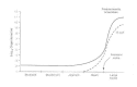
Figure 95-1
Concentration of the bacterial flora in regions of the gastrointestinal tract.
The character of the bacterial flora changes not only along the length of the gastrointestinal tract but also cross-sectionally with regard to the mucosal surface. Bacteria occupy the lumen, overlie the epithelial cells, and adhere to the mucosa. Penetration of bacteria through the mucosal surface is an abnormal event; pathogens such as Shigella, Salmonella, and Campylobacter invade in this way.
The same mechanisms that control the normal flora also protect the bowel from invasion by pathogens. Gastric acid in the stomach kills most organisms that are swallowed. Individuals with reduced or absent gastric acid have a high incidence of bacterial colonization in the upper small bowel and are more susceptible to bacterial diarrheal disease. Bile has antibacterial properties and thus may be another factor in controlling the flora. Forward propulsive motility (peristalsis) is a key element in suppressing the flora of the upper bowel. Finally, the microflora itself, by producing its own antibacterial substances (e.g., bacteriocins and fatty acids), stabilizes the normal populations and prevents implantation of pathogens.
Metabolic Activites of the Microflora
The metabolic capacities of the intestinal bacteria are extremely diverse. Bacterial enzymes can use as substrate virtually any compound in the intestinal lumen, whether taken orally or entering the intestine by secretion through the biliary tract or directly across the mucosa.
The Enterohepatic Circulation
Enzymes produced by intestinal bacteria play a central role in the enterohepatic circulation. Substances that undergo enterohepatic circulation are metabolized in the liver, excreted in the bile, and passed into the intestinal lumen, where they are reabsorbed across the intestinal mucosa and returned to the liver via the portal circulation. The enterohepatic circulation generally involves compounds that are conjugated in the liver to a polar group such as glucuronic acid, sulfate, taurine, glycine, or glutathione. Conjugation increases the solubility of the metabolite in bile, but the conjugated compounds are poorly absorbed by the intestinal mucosa. Enzymes produced by intestinal bacteria—such as ß-glucuronidase, sulfatase, and various glycosidases—deconjugate these compounds, releasing the parent compounds which are readily absorbed across the intestinal wall. Many endogenous compounds undergo enterohepatic circulation, including bilirubin, bile acids, cholesterol, estrogens, and metabolites of vitamin D. In addition, many drugs that are excreted by the liver, including digitalis, diethylstilbestrol, morphine, colchicine, rifampin, and chloramphenicol, enter this pathway.
Antibiotics block the enterohepatic circulation by suppressing the intestinal flora and thereby reducing the levels of deconjugating enzymes. If an antibiotic is given to a patient who is also taking a drug that undergoes enterohepatic circulation, the resulting depression of the enterohepatic circulation will increase the fecal excretion of the drug and thereby lower its plasma level and half life. For example, the blood levels and half life of the estrogen in birth control pills decrease when antibiotics are administered.
The Microflora and Nutrition
Enzymes produced by intestinal bacteria are important in the metabolism of several vitamins. The intestinal microflora synthesizes vitamin K, which is a necessary cofactor in the production of prothrombin and other blood clotting factors. Treatment with antibiotics, particularly in individuals eating a diet low in vitamin K, can result in low plasma prothrombin levels and a tendency to bleed. Intestinal bacteria also synthesize biotin, vitamin B12, folic acid, and thiamine.
The intestinal flora is capable of fermenting indigestible carbohydrates (dietary fiber) to short-chain fatty acids such as acetate, propionate, and butyrate. The major source of such fermentable carbohydrate in the human colon is plant cell wall polysaccharides such as pectins, cellulose, and hemicellulose. The acids produced from these fiber substrates by bacteria can be an important energy source for the host.
Some people are deficient in intestinal lactase, the mucosal enzyme responsible for hydrolyzing the disaccharide lactose in milk. In these individuals, lactose is not adequately digested and absorbed in the intestine. Lactose that reaches the large bowel undergoes vigorous bacterial fermentation. The result can be distention, flatus, and diarrhea.
The Intestinal Microflora and Infection
Protective Activities of the Flora
Like other complex ecosystems, the intestinal microflora is relatively stable over time, maintaining roughly constant numbers and types of bacteria in each area of the bowel. The stability of normal flora both discourages infection by exogenous pathogens and prevents overgrowth of potentially pathogenic members. New organisms that enter the system in contaminated food or water generally are suppressed by the established flora. This suppression is related to production by members of the resident flora of antimicrobial substances such as bacteriocins or short-chain fatty acids, which inhibit the growth of alien microorganisms. Antibiotics that kill off part of the intestinal flora can upset its balance and may open the door to infection or pathologic overgrowth.
The pathogenesis of Salmonella food poisoning illustrates this phenomenon. Normal individuals are quite resistant to Salmonella, and a large oral inoculum is required to initiate infection. If the intestinal flora is suppressed by antibiotics, however, the individual becomes much more susceptible and can be infected by a relatively small inoculum.
Diseases Caused by Overgrowth of Potential Pathogens
The normal intestinal flora includes small populations of organisms that cause disease if they overgrow. For example, overgrowth of Clostridium difficile produces severe inflammation of the colon with diarrhea (pseudomembranous colitis). Administration of antibiotics initiates the process by suppressing the normal flora.
Peritonitis
Bacteria from the intestinal flora are the prime cause of infection in the peritoneal cavity when the normal barriers of the intestinal wall are violated. The intestinal wall can be perforated by trauma (knife wounds, gunshot wounds, blunt trauma), by disease (appendicitis, penetrating intestinal cancers), or by surgical procedures. Once the mucosal barrier is breached, bacteria penetrate through the intestinal wall into the normally sterile peritoneal cavity and its surrounding structures. Poor circulation, reduced oxygen supply, and dead tissue in the vicinity of the perforation promote the formation of an abscess and particularly favor the growth of anaerobic bacteria. Cultures of a peritoneal abscess generally yield several types of bacteria from the intestinal microflora, particularly species of Bacteroides, Clostridium, and Peptostreptococcus and E coli.
Bacterial Diarrheas
Enterotoxin-Mediated Diarrheal Diseases
Several enterotoxin-producing bacteria cause diarrheal diseases (Table 95-1). The diarrheal disease caused by Vibrio cholerae and enterotoxigenic strains of E coli has three main characteristics. First, there is intestinal fluid loss that is related to the action of an enterotoxin on the small bowel epithelial cells. Second, the organism itself does not invade the mucosal surface; rather, it colonizes the upper small bowel, adhering to the epithelial cells and elaborating the enterotoxin. The mucosal architecture remains intact with no evidence of cellular destruction. Bacteremia does not occur. Third, the fecal effluent is watery and often voluminous, so that the diarrhea can result in clinical dehydration. The fluid originates in the upper small bowel. where the enterotoxin is most active.
Table 95-1
Toxin-Producing Bacteria Associated With Diarrheal Disease.
Cholera
The paradigm of the enterotoxigenic diarrheal diseases is cholera (see Ch. 24), in which stool volume can exceed 1 L/h, with daily fecal outputs of 15 to 20 L if the patient is kept hydrated. Cholera is caused by V cholerae, which is usually ingested in contaminated water. Vibrios that survive passage through the stomach colonize the surface of the small intestine, proliferate, and elaborate the enterotoxin. Cholera toxin acts via adenylate cyclase to stimulate secretion of water and electrolytes from the epithelial cells into the lumen of the gut. The duodenum and upper jejunum are more sensitive to the toxin than the ileum is. The colon is relatively insensitive to the toxin and may still absorb water and electrolytes normally. Thus, cholera is an “overflow diarrhea,” in which the large volumes of fluid produced in the upper intestine overwhelm the resorptive capacity of the lower bowel.
Cholera stool is described as resembling rice water—a clear fluid flecked with mucus—and is isotonic with plasma. Microscopy reveals no inflammatory cells in the fecal effluent; all that can be seen are small numbers of shed mucosal cells.
Enterotoxigenic E coli Diarrhea
Certain strains of E coli cause diarrheal disease by elaborating enterotoxins (see Ch. 25). These strains produce two types of enterotoxin. One, called heat-labile toxin, is similar in structure and in its mechanism of action to cholera toxin. The other, called heat-stable toxin, appears to act via guanylate cyclase. Enterotoxigenic E coli strains are the most common cause of travelers' diarrhea
Other Diarrhea-Causing Toxins
Many strains of Shigella produce an enterotoxin, called Shiga toxin, that causes secretion of fluid from the small intestine (see Ch. 22). Shiga toxin has a destructive, cytotoxic effect on the small-bowel epithelium, causing gross injury to the bowel surface. It does not activate adenylate cyclase. E coli 0157:H7, the organism associated with consumption of undercooked chopped meat, also produces a Shiga-like toxin; it causes bloody diarrhea and colitis. An organism that produces a different type of cytotoxin is Vibrio parahaemolyticus, a bacterium associated with seafood. Food-poisoning strains of Staphylococcus aureus and Clostridium perfringens both produce enterotoxins that are cytotoxic. The staphylococcal enterotoxin also has a direct effect on the vomiting center in the brain.
Gastrointestinal Disease Caused by Invasive Bacteria
Unlike the enterotoxigenic organisms, invasive bacteria exert their main impact on the host by causing gross destruction of the epithelial architecture; histologic findings include mucosal ulceration and an inflammatory reaction in the lamina propria. The principal pathogens in this group are Salmonella, Shigella, Campylobacter, invasive E coli, and Yersinia. The enteric viruses also invade intestinal epithelial cells, but the extent of mucosal destruction is considerably less than that caused by invasive bacterial pathogens.
Salmonella Enteritis
Salmonella species are a common cause of food poisoning. The main site of attack is the lower ileum, where the salmonellae cause mucosal ulceration. They rapidly make their way through the epithelial surface into the lamina propria and enter the lymphatics and bloodstream. At least two virulence factors are associated with intestinal infection: one responsible for mucosal invasion, and the other causing secretion of fluid and electrolytes into the bowel.
Shigella Dysentery
Shigella organisms cause bacillary dysentery, an invasive diarrheal disease of the lower bowel in which the stool contains an inflammatory exudate composed of polymorphonuclear leukocytes. The bacilli invade the epithelium of the colon and cause superficial ulceration. This invasive process depends on the presence of two virulence factors. The first mediates the initial penetration of the mucosal surface by destroying the brush border; the bacteria are subsequently engulfed by invagination of the plasma membrane. The second virulence factor allows the organism to multiply within the mucosal tissue. Mucosal ulceration results, accompanied by an intense inflammatory response in the lamina propria. The infection is usually restricted to the mucosa; lymph node involvement and bacteremia are uncommon.
Fluid Production in Invasive Diarrheal Diseases
The mechanism(s) by which the fluid that causes watery diarrhea is produced in the invasive diarrheal diseases is under debate. Three mechanisms have been proposed. First, Shigella and possibly Salmonella strains apparently produce an enterotoxin that stimulates the mucosa to secrete water and electrolytes. Second, there is evidence that invasive organisms stimulate prostaglandin synthesis at the site of inflammation and that the prostaglandins induce fluid secretion. In experimental animals, fluid secretion can be blocked by prostaglandin inhibitors such as indomethacin and aspirin. Third, some evidence suggests that damage to the colonic epithelium causes diarrhea by prevention of normal resorption of fluid.
Viral Diarrheas
Two viruses—rotavirus (see Ch. 63) and Calicivirus (Norwalk virus) (see Ch. 65)—have been identified as major enteric pathogens in humans. The rotaviruses are a very important cause of infantile diarrhea, which in undeveloped countries can be fatal. Adults may be infected and shed virus, but clinical disease appears almost exclusively in children younger than 2 years. Calicivirus, in contrast, can produce gastroenteritis in all age groups and is a cause of major epidemics. The initial lesion forms in the proximal small bowel. The mucosal architecture is damaged, with shortening of the villi and hyperplasia of the crypts. An inflammatory exudate then appears in the lamina propria.
The mechanisms responsible for fluid secretion in viral diarrheas have not been elucidated. It is known that infection with Calicivirus can produce steatorrhea and xylose malabsorption and causes direct damage to brush border enzymes. The activity of adenylate cyclase in the epithelial cells is not altered in the acute illness.
Parasitic Diarrheas
Several species of protozoa and helminths can cause diarrheal disease. Some of these infections can be acquired in the United States, although exposure to enteric parasites is far more common in tropical and developing countries. Some of the more common causes of parasitic diarrhea are Entamoeba histolytica, Giardia lamblia, Strongyloides stercoralis, and the intestinal flukes.
Clinical Diagnosis of Diarrheal Disease
An understanding of pathophysiology can be used to make a presumptive diagnosis in patients with infectious diarrhea (Table 95-2). Perhaps the most convenient approach is to separate pathogens that involve the small intestine from those that attack the large bowel. Enterotoxigenic bacteria (E coli, V cholerae), viruses, and the parasite Giardia are examples of small-bowel pathogens. These organisms produce watery diarrhea, which may lead to dehydration. Abdominal pain, although often diffuse and poorly defined, is generally periumbilical. Microscopic examination of the stool fails to reveal formed cellular elements such as erythrocytes and leukocytes.
Table 95-2
Clinical Features of Diarrheal Diseases.
The large-bowel pathogens (the major ones being Shigella and Campylobacter) are invasive organisms and cause the clinical syndrome known as dysentery. Involvement of the colon is strongly suggested by the characteristic rectal pain known as tenesmus. Although the fecal effluent may be watery at first, by the second or third day of illness the stool is scanty and often bloody or mucoid. Microscopic examination almost invariably reveals abundant erythrocytes and leukocytes. Proctoscopy shows a diffusely ulcerated, hemorrhagic, and friable colonic mucosa.
Salmonella food poisoning does not fit into this simple scheme, because the disease can display features typical of both small- and large-bowel disease. The organism is invasive for the mucosa of the small intestine, particularly the lower ileum, and can cause voluminous fluid secretion. In additional, septicemia and metastatic spread of the pathogen to other organs sometimes occur.
References
- Finegold S (ed): Centennial symposium on anaerobes: A memorial to Andre' Veillon. Clin Infect Dis 18:5–245, 1994 . [PubMed: 8086570]
- Goldin BR, Lichtenstein AH, Gorbach SL: The role of the intestinal flora. p. 500. In Shils ME, Young VR (eds): Modern Nutrition in Health and Disease. Lea & Febriger, Philadelphia, 1994 .
- Gorbach SL: Infectious diarrhea and bacterial food poisoning. p. 1128 In Sleisenger MH, Fordtran JS (eds): Gastrointestinal Diseases. WB Saunders, Philadelphia, 1993.
- Simon GL, Gorbach SL: Normal alimentary tract microflora. p. 53. In Blaser MJ, Smith PD, Ravdin JI, Greenberg HB, Guerrant RL (eds): Infections of the Gastrointestinal Tract, Raven Press, New York, 1995 .
- PubMedLinks to PubMed
- Review Microflora of the gastrointestinal tract: a review.[Methods Mol Biol. 2004]Review Microflora of the gastrointestinal tract: a review.Hao WL, Lee YK. Methods Mol Biol. 2004; 268:491-502.
- Review The normal human anaerobic microflora.[Scand J Infect Dis Suppl. 1982]Review The normal human anaerobic microflora.Evaldson G, Heimdahl A, Kager L, Nord CE. Scand J Infect Dis Suppl. 1982; 35:9-15.
- [Bacterial ecology of the digestive tract and defense of the body].[Ann Gastroenterol Hepatol (Par...][Bacterial ecology of the digestive tract and defense of the body].Rambaud JC. Ann Gastroenterol Hepatol (Paris). 1992 Nov-Dec; 28(6-7):263-6.
- Review Gastrointestinal microflora in mammalian nutrition.[Annu Rev Nutr. 1986]Review Gastrointestinal microflora in mammalian nutrition.Savage DC. Annu Rev Nutr. 1986; 6:155-78.
- Review Review article: gut flora and inflammatory bowel disease.[Aliment Pharmacol Ther. 2004]Review Review article: gut flora and inflammatory bowel disease.Marteau P, Lepage P, Mangin I, Suau A, Doré J, Pochart P, Seksik P. Aliment Pharmacol Ther. 2004 Oct; 20 Suppl 4:18-23.
- Microbiology of the Gastrointestinal Tract - Medical MicrobiologyMicrobiology of the Gastrointestinal Tract - Medical Microbiology
Your browsing activity is empty.
Activity recording is turned off.
See more...