Continuing Education Activity
Acromioclavicular joint injuries account for more than forty percent of all shoulder injuries. Mild injuries are not associated with any significant morbidity, but severe injuries can lead to significant loss of strength and function of the shoulder. Acromioclavicular injuries may be associated with a fractured clavicle, impingement syndromes, and more rarely neurovascular insults. This activity reviews the evaluation and management of this condition and highlights the role of the interprofessional team in caring for patients effected by this acromioclavicular joint injuries.
Objectives:
Describe the most common mechanism of injury associated with acromioclavicular joint injury.
Review the evaluation of a patient with a suspected acromioclavicular injury.
Identify the difference in the management of patients with Rockwood Classification type I-II injuries from patients with Rockwood Classification type III-VI.
Outline strategies for the interprofessional care team to provide optimal management and counseling to improve outcomes for patients with acromioclavicular joint injuries.
Access free multiple choice questions on this topic.
Introduction
Injury to the acromioclavicular (AC) joint is a common injury among athletes and young individuals. Acromioclavicular joint injuries account for more than forty percent of all shoulder injuries. Mild injuries are not associated with any significant morbidity, but severe injuries can lead to significant loss of strength and function of the shoulder. Acromioclavicular injuries may be associated with a fractured clavicle, impingement syndromes, and more rarely neurovascular insults.
Etiology
The AC joint is a diarthrodial joint defined by the lateral process of the clavicle articulating with the acromion process as it projects anteriorly off the scapula. The joint is primarily stabilized by the acromioclavicular ligament, which is composed of an anterior, posterior, inferior, and superior component. Of note, the superior portion of the AC ligament is the most important component for the stability of the AC joint. Supporting structures include two coracoclavicular ligaments (trapezoid and conoid ligaments), which provide vertical stability, as well as the coracoacromial ligament. Mild injuries are not associated with any significant morbidity, but severe injuries can lead to considerable loss of strength and function of the shoulder. Acromioclavicular injuries may be associated with a fractured clavicle, impingement syndromes, and more rarely neurovascular insults.[1][2]
Epidemiology
AC injuries are frequently seen after sporting events, car accidents, falls from a bicycle, and other sports-related activities (e.g. skiing). AC joint injuries may account for as much as 40% of all shoulder injuries and nearly 10% of all injuries in collision sports such as football, lacrosse, and ice hockey.[3][4]
Pathophysiology
The most common mechanism of injury is direct trauma to the lateral aspect of the shoulder or acromion process with the arm in adduction. Falling on an outstretched hand or elbow may also lead to AC joint separation.
History and Physical
Patients with an AC joint injury typically present with anterosuperior shoulder pain and will describe a mechanism of injury of blunt trauma to the abducted shoulder or landing on an outstretched arm, suggestive of this type of injury. They may describe pain radiating to the neck or shoulder, which is often worse with movement or when they try to sleep on the affected shoulder. On examination, the clinician may observe swelling, bruising, or a deformity of the AC joint, depending on the degree of injury. The patient will be tender at that location. They may have a restriction in the active and passive range of motion secondary to pain. "Piano key sign" may be seen, with an elevation of the clavicle that rebounds after inferior compression. Finally, It is essential to evaluate the entire clavicle for possible fracture or sternoclavicular injury as well as perform a full neurovascular exam on the affected extremity.
Evaluation
Standard X-rays are adequate to make a diagnosis of acromioclavicular joint injury and should be used to evaluate for other causes of traumatic shoulder pain. AC joint injuries may not always be evident on regular radiographic views (anteroposterior [AP], lateral). Additional views include the Zanca view; an AP view performed by tilting the beam 10 to 15 degrees cranial, as well as bilateral AP views to compare displacement to the contralateral shoulder. Weighted stress views may be useful to evaluate the displacement of the joint when the diagnosis is uncertain on standard AP views. If there is continued uncertainty in diagnosis, the provider may also consider ultrasound or MRI for further diagnostic evaluation.[5]
Consider the Rockwood Staging System outlined below on the evaluation of radiographs, especially when compared to the contralateral shoulder, which will be important for guiding treatment. Identification of the coracoclavicular interspace distance compared to the contralateral view will help to guide treatment options in the majority of cases. An example of the appropriate method for measuring coracoclavicular interspace distance appears below.
Treatment / Management
Acromioclavicular joint injuries follow the Rockwood classification system of type I to type VI. Type I is referred to as a sprain of the acromioclavicular ligaments only and demonstrates no radiographic displacement. Type II involves tearing of the acromioclavicular ligament and sprain of the coracoclavicular ligament with less than 25% increase in the coracoclavicular interspace or with the clavicle elevated but not superior to the border of the acromion. Type I and II sprains are managed non-operatively with a sling, analgesia, ice, and physical therapy. Type III AC joint separation involves tearing of both the acromioclavicular ligament and coracoclavicular ligaments resulting in clavicle elevation above the border of the acromion with a 25 to 100% increased coracoclavicular distance on x-ray compared to the contralateral side. Type III injuries are frequently managed non-operatively similar to type I and II; however, if the displacement is greater than 75%; the patient is a laborer, elite athlete, or concerned about cosmesis; or is not improving with conservative management, then surgical intervention may be considered. Posterior displacement of the distal clavicle into the trapezius defines type IV injuries. Superior displacement of the distal clavicle by more than 100% compared to contralateral defines type V injuries. Type VI is rare and is defined as an inferolateral displacement in a subacromial or subcoracoid displacement behind the coracobrachialis or biceps tendon. Type IV through VI injuries are typically managed surgically, and warrants referral to an orthopedic surgeon.[1][3][6]
Management of Acute Acromioclavicular Joint Disruption
If the AC joint injury presents within six weeks, it is considered acute. The main goal of treatment is acromioclavicular joint stabilization. Following techniques are used for stabilization and reduction of AC joint.[7]
Hook Plate
In the setting of Rockwood type Types III and V injuries, a distal clavicle hook plate may be employed to restore the alignment of the acromioclavicular joint. The "hook" portion of the hook plate is placed inferior to the acromion, with superior plating on the clavicle. While this does provide excellent reduction of the joint, the plate must be removed in a subsequent surgery to prevent subacromial irritation, subacromial impingement, acromion osteolysis, and iatrogenic damage to the rotator cuff.[8] Kienast et al. reported 84 % good and excellent results (Taft score) in type III–V AC disruption with a hook plate.[9] A superior approach is used for plate fixation. The hook is placed posteriorly under the acromion and screws are placed after the reduction of the AC joint.[7]
Bosworth Screw
All types of AC joint injuries can be treated with the Bosworth screw (CC screw fixation), which has outstanding results and is very cost-effective. Bosworth screw along with acromioclavicular (AC) and coracoclavicular (CC) ligament reconstruction provide excellent long-term outcomes. A 6.5mm screw is placed from the clavicle to the coracoid through a mini-open incision or percutaneously. Early screw removal (usually 8 weeks) is necessary to prevent fatigue failure and crew breakage.[10][11]
Tension Band Wiring
Tucek et al. compared tension band wiring versus hook plate in 80 patients with type 5 AC joint disruption. Tension band wiring(TBW) provides comparable clinico-radiological results however hook plate allows an earlier range of motion and has a reduced rate of complications. TBW should be removed after 8 weeks to prevent wire malposition, migration, and breakage.[12]
EndoButton
A suitable treatment option for acromioclavicular dislocation is coracoclavicular ligament reconstruction utilizing an endobutton, which has the benefits of solid fixation and an early functional range of motion. [13] Double or triple Ethibond no.5 loops are used with titanium endobuttons from clavicle to coracoid. Continuous and double loops minimize the risk of loop-knot slippage as compared to a single suspension system.[14]
Management of Chronic Acromioclavicular Joint Disruption
There are three main goals of treatment
AC joint debridement
Ligament reconstruction ( acromioclavicular/ coracoclavicular)
Stable fixation
Following procedures are used for chronic AC joint disruptions.
Modified Weaver-Dunn procedure
An incision is made from the acromioclavicular joint the tip of the coracoid. After raising skin flaps, the acromioclavicular joint is debrided and a distal 1cm of the clavicle is excised. The coracoacromial ligament is transferred to the distal end of the clavicle along with a small piece of the acromion. The modification in the original procedure is done in the form of added fixation with a screw or plate. [15][16]
Mazzocca Technique
The incision extends from the tip of the coracoid to 2 cm posterior to the AC joint. The distal 1 cm of the clavicle is excised after AC joint debridement. A semitendinosus autograft is prepared and looped around the coracoid. The anatomical reconstruction is done by making bony tunnels in the shaft of the clavicle at foot print of the conoid and trapezoid ligament.[17]
Neviaser Technique
The coracoacromial ligament is mobilized from the coracoid along with a small bony piece from its attachment site. K-wires are used to anchor the ligament to the distal end of the clavicle.[18]
Rockwood Procedure
The original procedure includes the transfer of coracoacromial ligament from the acromion to the distal end of the clavicle. The bony tunnels are made through the lateral end of the clavicle to anchor this CA ligament. Stable reduction is provided by a Bosworth screw to secure this ligament reconstruction.
Docking Technique
The coracoacromial ligament is transferred from the acromion into the distal end of the clavicle. The docking procedure involves tensioning the CA ligament into the distal clavicle's medullary canal by using 2 nonabsorbable sutures.[19]
Mumford Procedure
This procedure is required for chronic type 1 & 2 AC joint disruptions in which ligaments are intact. The distal 2.5 cm of the clavicle is excised to reduce joint pain. The soft tissue and periosteum are pilcated over the lateral end of the clavicle.[20]
Staging
Rockwood Classification of AC Joint Injuries (acromioclavicular [AC], coracoclavicular [CC]).[21]
I: AC ligament sprain; CC ligament intact; no radiographic abnormalities
II: AC ligament is torn; CC ligament sprain; the clavicle has elevated but is not superior to the border of the acromion, or exhibits a less than 25% increase in the CC interspace compared to the contralateral
III: AC and CC ligaments are torn; clavicle has elevated above the border of the acromion, or there is an increase of 25% to 100% in the CC interspace compared to the contralateral
IV: AC and CC ligaments are torn; posterior displacement of the distal clavicle into the trapezius
V: AC and CC ligaments are torn; superior displacement of the distal clavicle by more than 100% in the CC interspace compared to the contralateral
VI: AC and CC ligaments are torn; inferolateral displacement in a subacromial or subcoracoid displacement behind the coracobrachialis or biceps tendon
The Rockwood classification remains the gold standard for driving treatment decisions and is an excellent tool in the management of acromioclavicular joint injuries.
Prognosis
The prognosis for AC joint injuries is generally favorable. Most injuries receive non-operative management, and individuals typically regain functional motion by six weeks and return to normal activity by 12 weeks. Surgically managed injuries have a more extended recovery timeframe, including immobilization for six weeks and a gradual return to full activity around six months.[22][23][24]
Complications
The most frequently encountered complication of AC joint separation is residual joint pain affecting anywhere from 30% to 50% of individuals. AC joint arthritis is also a known complication and is more common with surgical management. Hardware irritation, infection, adhesive capsulitis, coracoid, and clavicular fractures are common complications following fixation. Hook plate leads to acromion irritation, subacromial impingement, and osteolysis. [25]
Postoperative and Rehabilitation Care
1st-6th Weeks[26]
An arm sling is applied
Ice packs are applied to shoulder
Elbow and wrist mobilization is started
Auxiliary hygiene is maintained
Adequate analgesia is provided
6th-12th Weeks
Limit abduction above 90 degrees
Wean off polysling and start pendulum exercises
Passive and then active range of motion exercises are started
Start isometric and then strengthening exercises of rotator cuff
Elbow and wrist mobilization is continued
> 12weeks
The patient is allowed to return to sports after 6 months.[27]
Consultations
Type I and II AC joint separations do not necessarily require an evaluation by a specialist; however, type III through VI should have an evaluation by an orthopedic surgeon or sports medicine physician.
Deterrence and Patient Education
Injuries of the acromioclavicular joint are common causes of shoulder pain and frequently occur in athletes and the setting of traumatic injury. Patients should seek an evaluation from a trained orthopedic or sports management physician. Early management with sling immobilization, ice, rest, and anti-inflammatory medications is generally an appropriate treatment for these injuries. Patients can generally return to normal activities after they have been evaluated and cleared by their treating physician.
Pearls and Other Issues
The position of the AC joint makes it vulnerable to direct trauma to the superior or lateral aspect of the shoulder.
In an isolated injury, patients typically have pain and swelling over the joint with or without a deformity depending on the degree of injury.
All patients should receive a radiographic evaluation of the affected shoulder, and if the diagnosis is in question, obtain radiographs of the contralateral AC joint as well.
Type I and II injuries occur twice as frequently as type III to VI injuries.
Most injuries can have management non-operatively. If uncertainty exists or it is a Rockwell grade III or greater, an orthopedic physician should be urgently consulted.
Non-operative management includes rest, protection with a sling, ice, NSAIDs, and physical therapy.
Enhancing Healthcare Team Outcomes
AC joint injuries commonly present to the emergency department. The mild injuries are usually managed conservatively, but the severe injuries may require surgery; hence it is crucial to consult an orthopedic surgeon when arriving at the diagnosis, to grade the injury. For mild injuries, the nurse practitioner, emergency department physician, and primary caregiver should recommend physical therapy, once the acute pain has resolved. Pharmacists and nurses should work with the team to educate the patient and family and assist in pain control. Those wishing to return to sports should first consult with the orthopedic surgeon.[28]
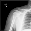
Shoulder Radiograph, Acromioclavicular (AC) Joint Separation With Injury Type III/IV Contributed by Scott Dulebohn, MD
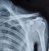
Acromioclavicular joint disruption Dr. Muhammad Taqi
References
- 1.
Gowd AK, Liu JN, Cabarcas BC, Cvetanovich GL, Garcia GH, Manderle BJ, Verma NN. Current Concepts in the Operative Management of Acromioclavicular Dislocations: A Systematic Review and Meta-analysis of Operative Techniques.
Am J Sports Med. 2019 Sep;47(11):2745-2758. [
PubMed: 30272997]
- 2.
Hyland S, Charlick M, Varacallo M.
StatPearls [Internet]. StatPearls Publishing; Treasure Island (FL): Jul 24, 2023. Anatomy, Shoulder and Upper Limb, Clavicle. [
PubMed: 30252246]
- 3.
Sirin E, Aydin N, Mert Topkar O. Acromioclavicular joint injuries: diagnosis, classification and ligamentoplasty procedures.
EFORT Open Rev. 2018 Jul;3(7):426-433. [
PMC free article: PMC6129955] [
PubMed: 30233818]
- 4.
Ruiz Ibán MA, Sarasquete J, Gil de Rozas M, Costa P, Tovío JD, Carpinteiro E, Hachem AI, Perez España M, Asenjo Gismero C, Diaz Heredia J, García Navlet M. Low prevalence of relevant associated articular lesions in patients with acute III-VI acromioclavicular joint injuries.
Knee Surg Sports Traumatol Arthrosc. 2019 Dec;27(12):3741-3746. [
PubMed: 30097689]
- 5.
Ernberg LA, Potter HG. Radiographic evaluation of the acromioclavicular and sternoclavicular joints.
Clin Sports Med. 2003 Apr;22(2):255-75. [
PubMed: 12825529]
- 6.
Hashiguchi H, Iwashita S, Abe K, Sonoki K, Yoneda M, Takai S. Arthroscopic Coracoclavicular Ligament Reconstruction for Acromioclavicular Joint Dislocation.
J Nippon Med Sch. 2018;85(3):166-171. [
PubMed: 30135343]
- 7.
Martetschläger F, Kraus N, Scheibel M, Streich J, Venjakob A, Maier D. The Diagnosis and Treatment of Acute Dislocation of the Acromioclavicular Joint.
Dtsch Arztebl Int. 2019 Feb 08;116(6):89-95. [
PMC free article: PMC6435864] [
PubMed: 30892184]
- 8.
Hung LK, Su KC, Lu WH, Lee CH. Biomechanical analysis of clavicle hook plate implantation with different hook angles in the acromioclavicular joint.
Int Orthop. 2017 Aug;41(8):1663-1669. [
PubMed: 28097386]
- 9.
Kienast B, Thietje R, Queitsch C, Gille J, Schulz AP, Meiners J. Mid-term results after operative treatment of rockwood grade III-V acromioclavicular joint dislocations with an AC-hook-plate.
Eur J Med Res. 2011 Feb 24;16(2):52-6. [
PMC free article: PMC3353421] [
PubMed: 21463981]
- 10.
Lowe GP, Fogarty MJ. Acute acromioclavicular joint dislocation: results of operative treatment with the Bosworth screw.
Aust N Z J Surg. 1977 Oct;47(5):664-7. [
PubMed: 273410]
- 11.
Jeong JY, Chun YM. Treatment of acute high-grade acromioclavicular joint dislocation.
Clin Shoulder Elb. 2020 Sep;23(3):159-165. [
PMC free article: PMC7714286] [
PubMed: 33330252]
- 12.
Tuček M, Chochola A, Vaněček V, Bušková K. [Surgical treatment of acromioclavicular dislocation: Tension band wiring versus hook plate].
Rozhl Chir. 2015 Oct;94(10):437-44. [
PubMed: 26556021]
- 13.
Chen MM, Ye XY, Ni YP, Mou ZF, Huang LP. [Application of endobutton in the treatment of acute acromioclavicular joint dislocation].
Zhongguo Gu Shang. 2011 Mar;24(3):189-91. [
PubMed: 21485561]
- 14.
Lim YW. Triple endobutton technique in acromioclavicular joint reduction and reconstruction.
Ann Acad Med Singap. 2008 Apr;37(4):294-9. [
PubMed: 18461213]
- 15.
Galasso O, Tarducci L, De Benedetto M, Orlando N, Mercurio M, Gasparini G, Castricini R. Modified Weaver-Dunn Procedure for Type 3 Acromioclavicular Joint Dislocation: Functional and Radiological Outcomes.
Orthop J Sports Med. 2020 Mar;8(3):2325967120905022. [
PMC free article: PMC7065288] [
PubMed: 32215276]
- 16.
Pavlik A, Csépai D, Hidas P. Surgical treatment of chronic acromioclavicular joint dislocation by modified Weaver-Dunn procedure.
Knee Surg Sports Traumatol Arthrosc. 2001 Sep;9(5):307-12. [
PubMed: 11685364]
- 17.
Mazzocca AD, Arciero RA, Bicos J. Evaluation and treatment of acromioclavicular joint injuries.
Am J Sports Med. 2007 Feb;35(2):316-29. [
PubMed: 17251175]
- 18.
Sood A, Wallwork N, Bain GI. Clinical results of coracoacromial ligament transfer in acromioclavicular dislocations: A review of published literature.
Int J Shoulder Surg. 2008 Jan;2(1):13-21. [
PMC free article: PMC3022141] [
PubMed: 21264150]
- 19.
Millett PJ, Braun S, Gobezie R, Pacheco IH. Acromioclavicular joint reconstruction with coracoacromial ligament transfer using the docking technique.
BMC Musculoskelet Disord. 2009 Jan 14;10:6. [
PMC free article: PMC2637828] [
PubMed: 19144190]
- 20.
Snyder SJ, Banas MP, Karzel RP. The arthroscopic Mumford procedure: an analysis of results.
Arthroscopy. 1995 Apr;11(2):157-64. [
PubMed: 7794427]
- 21.
Granville-Chapman J, Torrance E, Rashid A, Funk L. The Rockwood classification in acute acromioclavicular joint injury does not correlate with symptoms.
J Orthop Surg (Hong Kong). 2018 May-Aug;26(2):2309499018777886. [
PubMed: 29792117]
- 22.
Stein T, Müller D, Blank M, Reinig Y, Saier T, Hoffmann R, Welsch F, Schweigkofler U. Stabilization of Acute High-Grade Acromioclavicular Joint Separation: A Prospective Assessment of the Clavicular Hook Plate Versus the Double Double-Button Suture Procedure.
Am J Sports Med. 2018 Sep;46(11):2725-2734. [
PubMed: 30106600]
- 23.
Byrne PA, Hopper GP, Wilson WT, Mackay GM. Acromioclavicular Joint Stabilisation Using the Internal Brace Principle.
Surg Technol Int. 2018 Nov 11;33:294-298. [
PubMed: 30029288]
- 24.
López-Alameda S, Fernández-Santás T, García-Villanueva A, Varillas-Delgado D, Garcia de Lucas F. Results of surgical treatment of acromioclavicular dislocations type III using modified Weaver Dunn technique.
Rev Esp Cir Ortop Traumatol (Engl Ed). 2018 Mar-Apr;62(2):93-99. [
PubMed: 29428418]
- 25.
Ma R, Smith PA, Smith MJ, Sherman SL, Flood D, Li X. Managing and recognizing complications after treatment of acromioclavicular joint repair or reconstruction.
Curr Rev Musculoskelet Med. 2015 Mar;8(1):75-82. [
PMC free article: PMC4596186] [
PubMed: 25663435]
- 26.
LeVasseur MR, Mancini MR, Berthold DP, Cusano A, McCann GP, Cote MP, Gomlinski G, Mazzocca AD. Acromioclavicular Joint Injuries: Effective Rehabilitation.
Open Access J Sports Med. 2021;12:73-85. [
PMC free article: PMC8169819] [
PubMed: 34093044]
- 27.
Cheema SG, Hermanns C, Coda RG, Tarakemeh A, Mullen SM, Schroeppel JP, Vopat BG, Mulcahey MK. Publicly Accessible Rehabilitation Protocols for Acromioclavicular Joint Reconstruction Are Widely Variable.
Arthrosc Sports Med Rehabil. 2021 Apr;3(2):e427-e433. [
PMC free article: PMC8129471] [
PubMed: 34027451]
- 28.
Müller D, Reinig Y, Hoffmann R, Blank M, Welsch F, Schweigkofler U, Stein T. Return to sport after acute acromioclavicular stabilization: a randomized control of double-suture-button system versus clavicular hook plate compared to uninjured shoulder sport athletes.
Knee Surg Sports Traumatol Arthrosc. 2018 Dec;26(12):3832-3847. [
PubMed: 29980805]
Disclosure: John Kiel declares no relevant financial relationships with ineligible companies.
Disclosure: Muhammad Taqi declares no relevant financial relationships with ineligible companies.
Disclosure: Kimberly Kaiser declares no relevant financial relationships with ineligible companies.