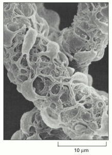
From: Blood Vessels and Endothelial Cells

Molecular Biology of the Cell. 4th edition.
Alberts B, Johnson A, Lewis J, et al.
New York: Garland Science; 2002.
Copyright © 2002, Bruce Alberts, Alexander Johnson, Julian
Lewis, Martin Raff, Keith Roberts, and Peter Walter; Copyright © 1983,
1989, 1994, Bruce Alberts, Dennis Bray, Julian Lewis, Martin Raff, Keith Roberts,
and James D. Watson .
NCBI Bookshelf. A service of the National Library of Medicine, National Institutes of Health.
