From: Blood Vessels and Endothelial Cells

NCBI Bookshelf. A service of the National Library of Medicine, National Institutes of Health.

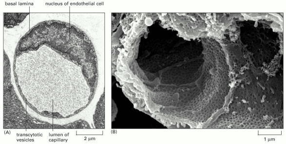
(A) Electron micrograph of a cross section of a small capillary in the pancreas. The wall is formed by a single endothelial cell surrounded by a basal lamina. Note the small “transcytotic” vesicles, which according to one theory provide transport of large molecules in and out of this type of capillary: materials are taken up into the vesicles by endocytosis at the luminal surface of the cell and discharged by exocytosis at the external surface, or vice versa. (B) Scanning electron micrograph of the interior of a capillary in a glomerulus of the kidney, where filtration of the blood occurs to produce urine. Here, as in the liver (see Figure 22-20), the endothelial cells are specialized to form a sieve-like structure, with fenestrae (“windows”), constructed rather like the pores in the nuclear envelope of eucaryotic cells, allowing water and most molecules to pass freely out of the bloodstream. (A, from R.P. Bolender, J. Cell Biol. 61:269–287, 1974. © The Rockefeller University Press; B, courtesy of Steve Gschmeissner and David Shima.)
From: Blood Vessels and Endothelial Cells

NCBI Bookshelf. A service of the National Library of Medicine, National Institutes of Health.